Key takeaways
- An Oxygenation Index (OI) greater than 25 indicates severe hypoxemia and is a critical indicator for ECMO evaluation.
- A pre/post-ductal SpO2 difference greater than 5-10% confirms right-to-left shunting via a patent ductus arteriosus in PPHN.
- NT-proBNP greater than 850 pg/mL demonstrates high sensitivity for predicting the need for inhaled nitric oxide (iNO) therapy.
- The standard initial dose of inhaled nitric oxide (iNO) is 20 ppm for term or near-term infants, with 5 to 10 ppm for preterm infants and 40-80 ppm for refractory cases.
- PPHN has 3 clinical phenotypes based on the site of resistance: Type 1 (pre-capillary), Type 2 (increased flow), and Type 3 (post-capillary, e.g., TAPVC or LV dysfunction).
References
- Chojnacka K, Singh Y, Gahlaut S, Blaz W, Jerzak A, Szczapa T. Persistent Pulmonary Hypertension of the Newborn: A Pragmatic Review of Pathophysiology, Diagnosis, and Advances in Management. Biomedicines 2025, 13, 2332.
- Mark C. Mammel (editor), Anton H. L. C. van Kaam (editor), Steven M. Donn (editor) – Manual of neonatal respiratory care. (2022), p. 628 — ECMO criterion.
Cover
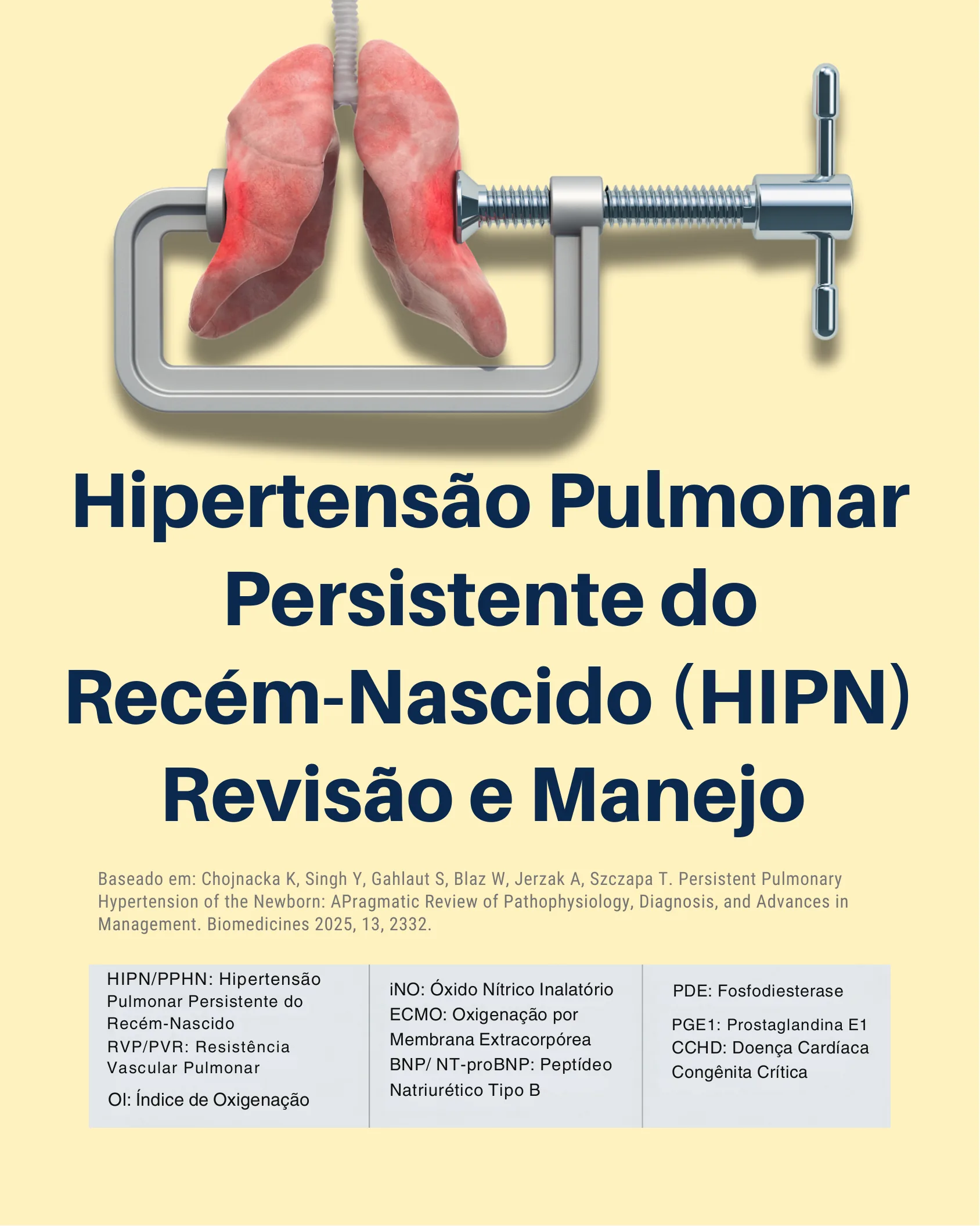
Persistent Pulmonary Hypertension of the Newborn (PPHN): Review and Management
Source: Chojnacka K, Singh Y, Gahlaut S, Blaz W, Jerzak A, Szczapa T. Persistent Pulmonary Hypertension of the Newborn: A Pragmatic Review of Pathophysiology, Diagnosis, and Advances in Management. Biomedicines 2025, 13, 2332.
- HIPN/PPHN: Hypertension Persistent Pulmonary Syndrome of the Newborn
- RVP/PVR: Resistance Pulmonary Vascular
- OI: Oxygenation Index
- iNO: Inhaled Nitric Oxide
- ECMO: Extracorporeal Membrane Oxygenation
- BNP/NT-proBNP: B-type natriuretic peptide
- PDE: Phosphodiesterase
- PGE1: Prostaglandina E1
- CCHD: Critical Congenital Heart Disease
The Collapse of the Fetal-Neonatal Transition
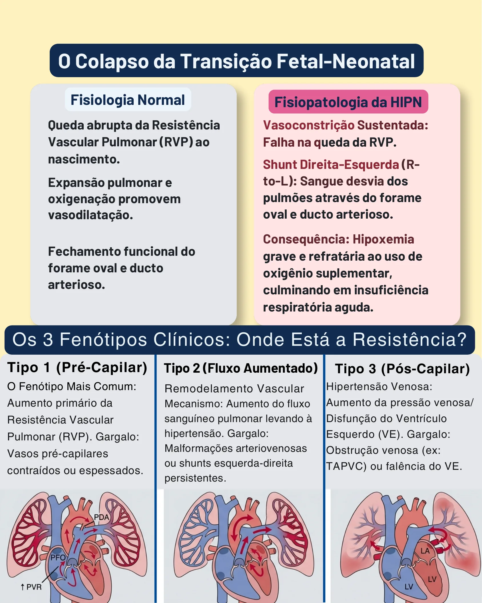
The Collapse of the Fetal-Neonatal Transition
Normal Physiology
- Abrupt drop in Pulmonary Vascular Resistance (PVR) at birth.
- Lung expansion and oxygenation promote vasodilation.
- Functional closure of the foramen ovale and ductus arteriosus.
The pathophysiology of HIPN
- Sustained Vasoconstriction: Failure to reduce pulmonary vascular resistance (PVR).
- Right-to-Left (R-to-L) Shunt: Blood is diverted away from the lungs through the foramen ovale and ductus arteriosus.
- Consequence: Hypoxemia severe and refractory to the use of supplemental oxygen, culminating in acute respiratory failure.
The 3 Clinical Phenotypes: Where is the Resistance?
Type 1 (Pre-Capillary)
Most Common Phenotype: Primary Increase in Pulmonary Vascular Resistance (PVR). Bottleneck: Constricted or thickened pre-capillary vessels.
Type 2 (Increased Flow)
Vascular Remodeling Mechanism: Increased pulmonary blood flow leading to hypertension. Bottleneck: Persistent arteriovenous malformations or left-to-right shunts.
Type 3 (Post-Capillary)
Venous Hypertension: Increased venous pressure/Left Ventricular (LV) Dysfunction. Bottleneck: Venous obstruction (e.g., TAPVC) or LV failure.
Etiology and Risk Factors
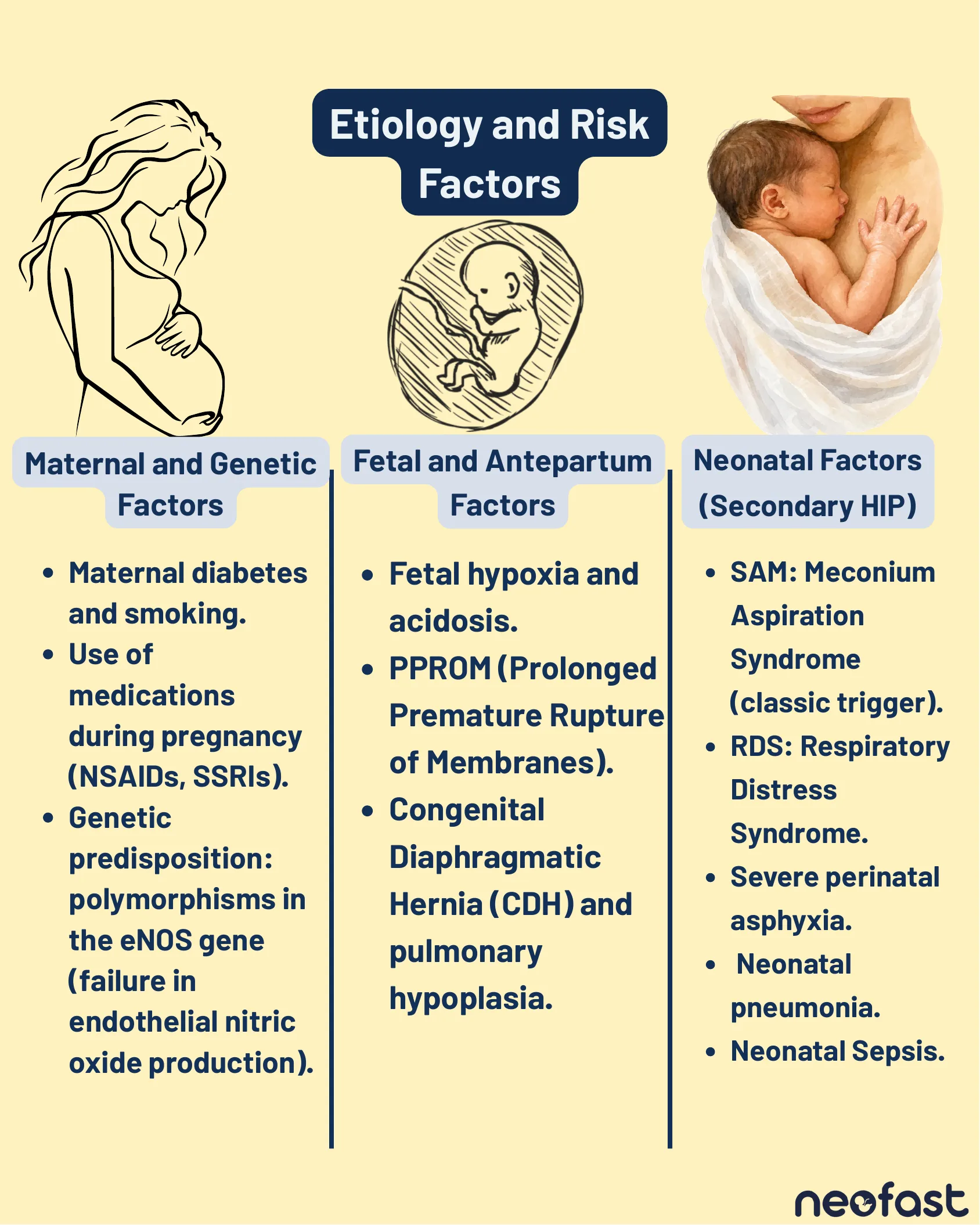
Etiology and Risk Factors
Maternal and Genetic Factors
- Maternal diabetes and smoking.
- Use of medications during pregnancy (NSAIDs, SSRIs).
- Genetic predisposition: polymorphisms in the eNOS gene (failure in endothelial nitric oxide production).
Fetal and Antepartum Factors
- Fetal hypoxia and acidosis.
- PPROM (Prolonged Premature Rupture of Membranes).
- Congenital Diaphragmatic Hernia (CDH) and pulmonary hypoplasia.
Neonatal Factors (Secondary HIP)
- SAM: Meconium Aspiration Syndrome (classic trigger).
- RDS: Respiratory Distress Syndrome.
- Severe perinatal asphyxia.
- Neonatal pneumonia.
- Neonatal Sepsis.
Etiology and Risk Factors
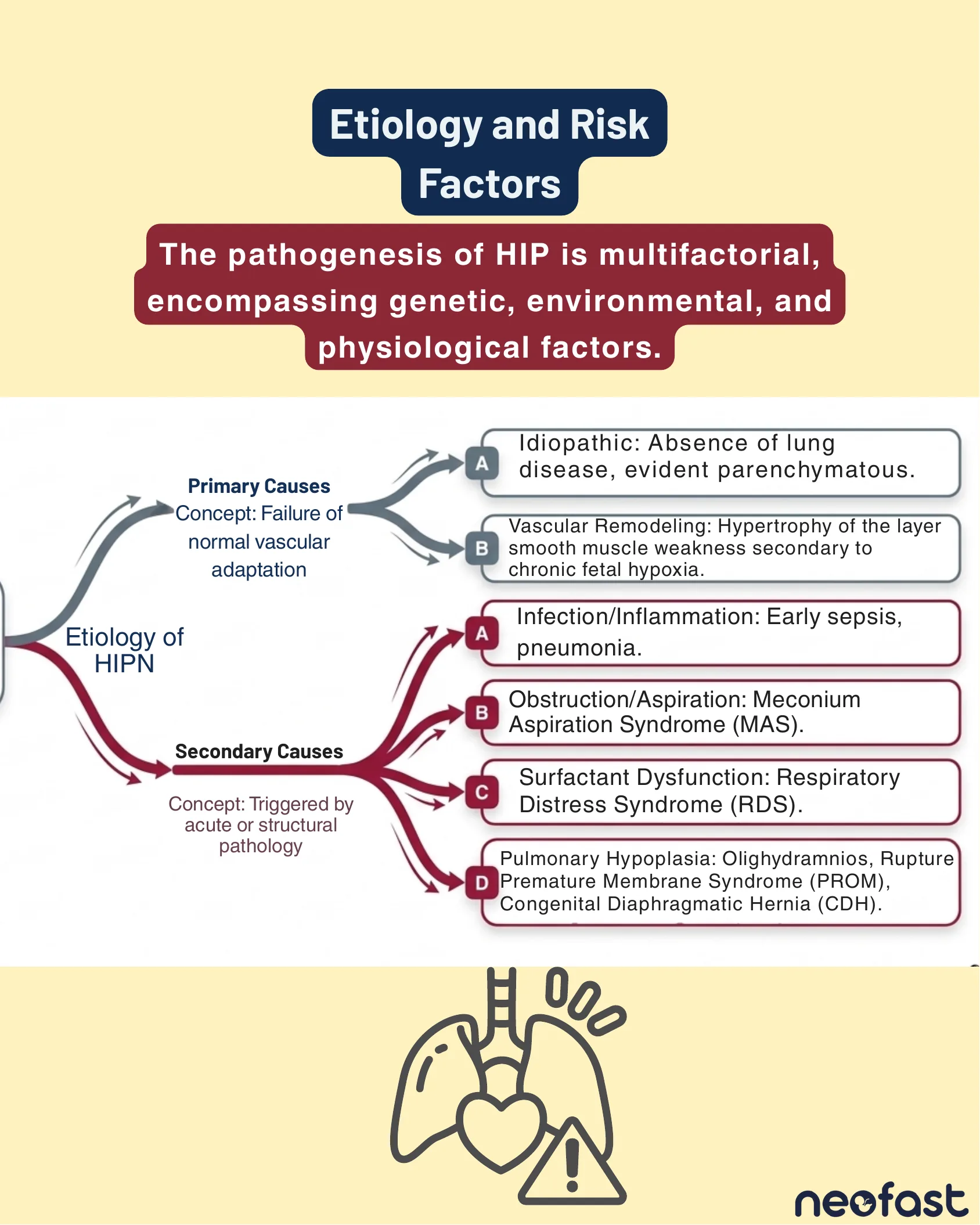
Etiology and Risk Factors
The pathogenesis of HIP is multifactorial, encompassing genetic, environmental, and physiological factors.
Primary Causes — Concept: Failure of normal vascular adaptation
- A. Idiopathic: Absence of lung disease, evident parenchymatous.
- B. Vascular Remodeling: Hypertrophy of the layer smooth muscle weakness secondary to chronic fetal hypoxia.
Secondary Causes — Concept: Triggered by acute or structural pathology
- A. Infection/Inflammation: Early sepsis, pneumonia.
- B. Obstruction/Aspiration: Meconium Aspiration Syndrome (MAS).
- C. Surfactant Dysfunction: Respiratory Distress Syndrome (RDS).
- D. Pulmonary Hypoplasia: Oligohydramnios, Rupture Premature Membrane Syndrome (PROM), Congenital Diaphragmatic Hernia (CDH).
Clinical Diagnosis
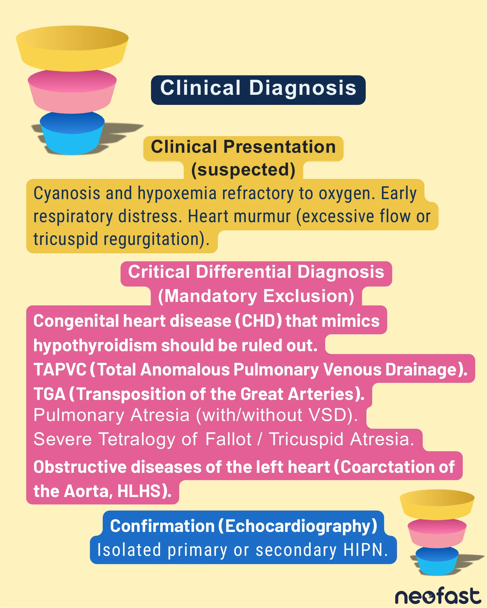
Clinical Diagnosis
Clinical Presentation (suspected)
Cyanosis and hypoxemia refractory to oxygen. Early respiratory distress. Heart murmur (excessive flow or tricuspid regurgitation).
Critical Differential Diagnosis (Mandatory Exclusion)
- Congenital heart disease (CHD) that mimics hypothyroidism should be ruled out.
- TAPVC (Total Anomalous Pulmonary Venous Drainage).
- TGA (Transposition of the Great Arteries).
- Pulmonary Atresia (with/without VSD).
- Severe Tetralogy of Fallot / Tricuspid Atresia.
- Obstructive diseases of the left heart (Coarctation of the Aorta, HLHS).
Confirmation (Echocardiography)
Isolated primary or secondary HIPN.
Clinical Diagnosis
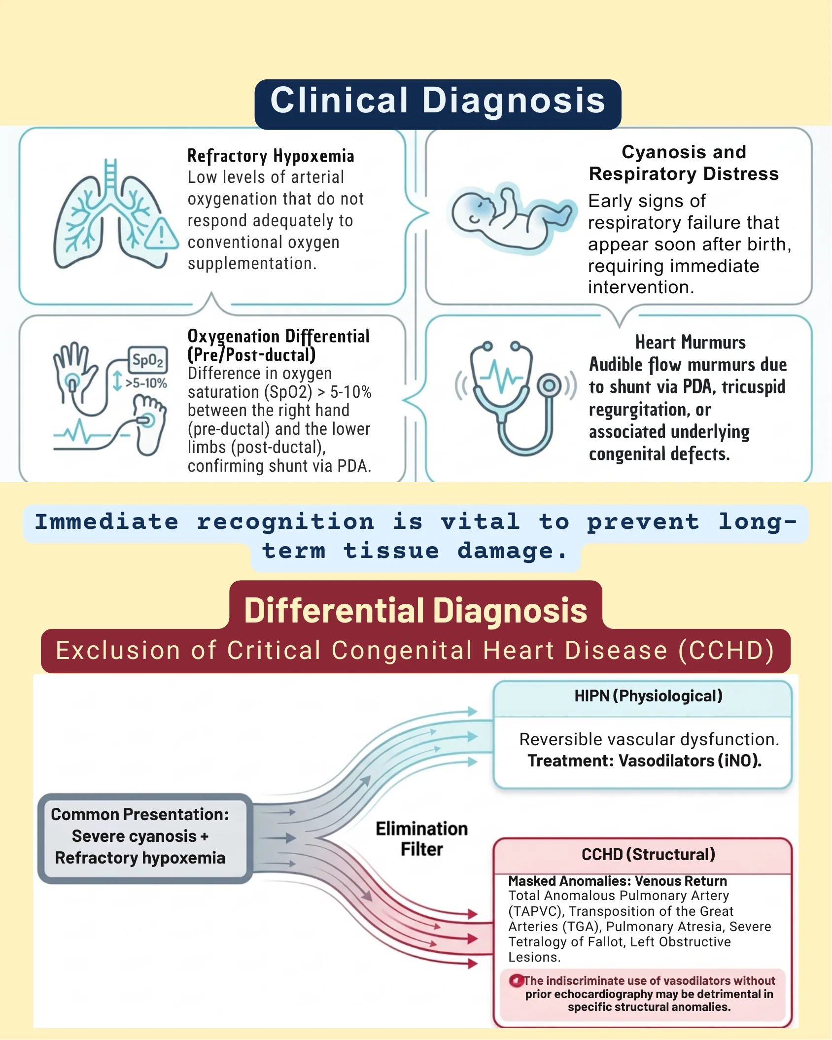
Clinical Diagnosis
Refractory Hypoxemia
Low levels of arterial oxygenation that do not respond adequately to conventional oxygen supplementation.
Cyanosis and Respiratory Distress
Early signs of respiratory failure that appear soon after birth, requiring immediate intervention.
Oxygenation Differential (Pre/Post-ductal)
Difference in oxygen saturation (SpO2) > 5-10% between the right hand (pre-ductal) and the lower limbs (post-ductal), confirming shunt via PDA.
Heart Murmurs
Audible flow murmurs due to shunt via PDA, tricuspid regurgitation, or associated underlying congenital defects.
Immediate recognition is vital to prevent long-term tissue damage.
Differential Diagnosis — Exclusion of Critical Congenital Heart Disease (CCHD)
Common Presentation: Severe cyanosis + Refractory hypoxemia → Elimination Filter →
- HIPN (Physiological): Reversible vascular dysfunction. Treatment: Vasodilators (iNO).
- CCHD (Structural): Masked Anomalies: Venous Return — Total Anomalous Pulmonary Artery (TAPVC), Transposition of the Great Arteries (TGA), Pulmonary Atresia, Severe Tetralogy of Fallot, Left Obstructive Lesions.
The indiscriminate use of vasodilators without prior echocardiography may be detrimental in specific structural anomalies.
Gold Standard: Bedside Echocardiography
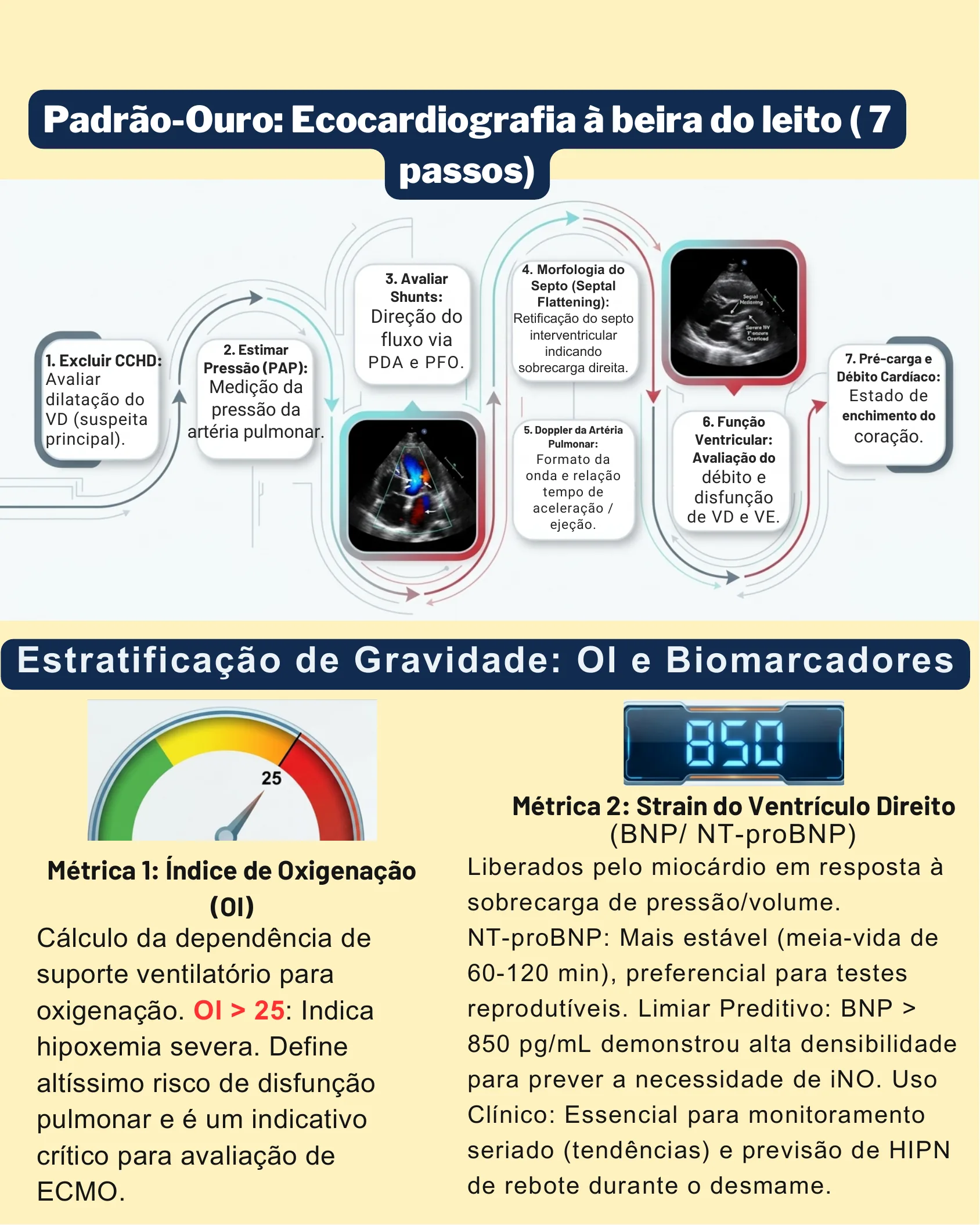
Gold Standard: Bedside Echocardiography (7 steps)
- Exclude CCHD: Assess right ventricular dilation (suspected principal).
- Estimate Pressure (PAP): Pulmonary artery pressure measurement.
- Evaluate Shunts: Direction of flow via PDA e PFO.
- Septal Morphology (Septal Flattening): Straightening of the interventricular septum indicating right ventricular overload.
- Pulmonary Artery Doppler: Waveform and acceleration/ejection time relationship.
- Ventricular Function: Evaluation of debt and dysfunction de RV e LV.
- Preload and Cardiac Output: State of filling of heart.
Gravity Stratification: OI and Biomarkers
Metric 1: Oxygenation Index (OI)
Calculation of dependence on ventilatory support for oxygenation. OI > 25: Indicates severe hypoxemia. Defines a very high risk of pulmonary dysfunction and is a critical indicator for ECMO evaluation.
Metric 2: Right Ventricular Strain (BNP/NT-proBNP)
Released by the myocardium in response to pressure/volume overload. NT-proBNP: More stable (half-life of 60-120 min), preferred for reproducible tests. Predictive Threshold: BNP > 850 pg/mL demonstrated high density for predicting the need for iNO. Clinical Use: Essential for serial monitoring (trends) and prediction of rebound HIP during weaning.
Severity Classification: Oxygenation Index (OI)
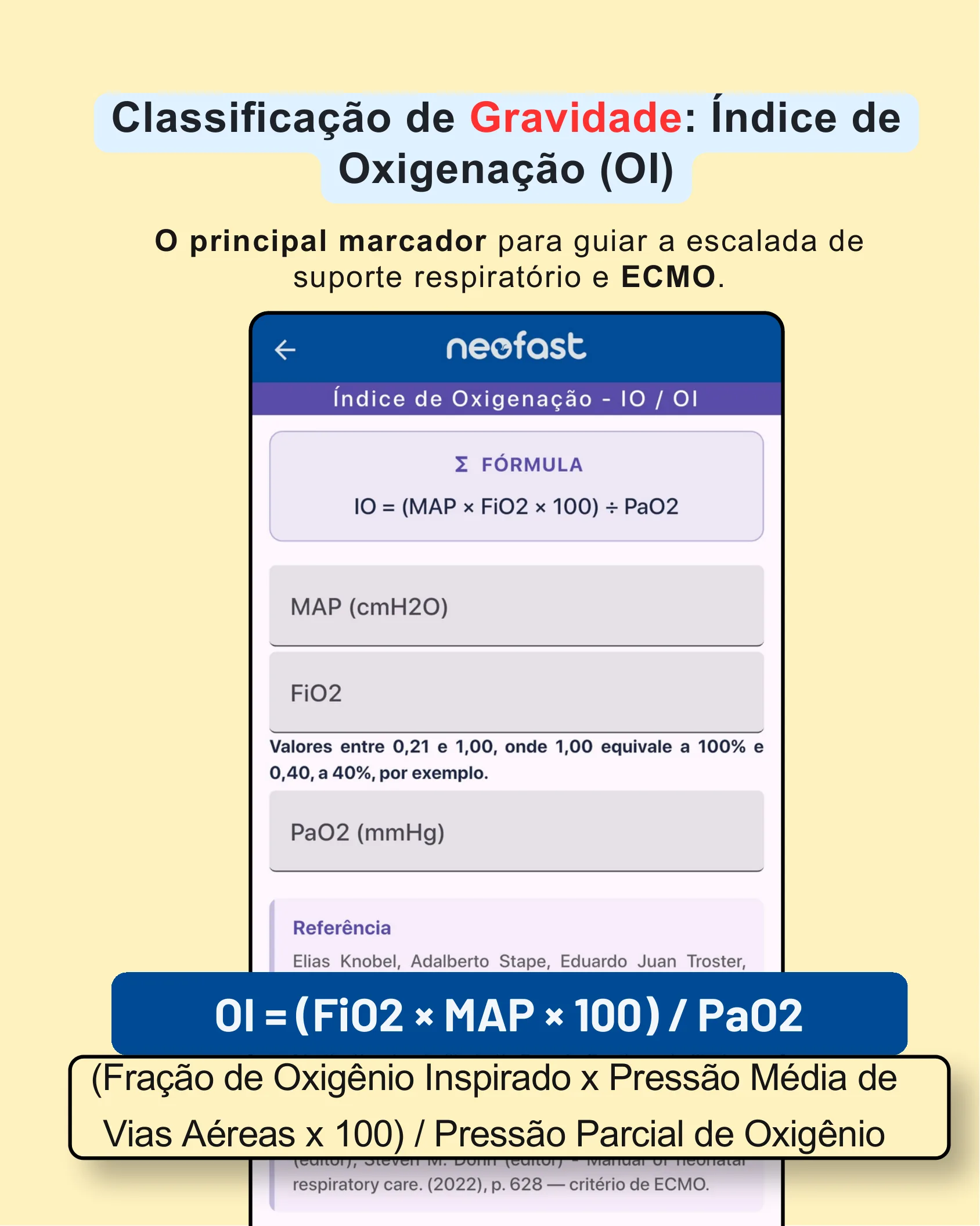
Severity Classification: Oxygenation Index (OI)
The primary marker to guide the escalation of respiratory support and ECMO.
Formula
OI = (MAP × FiO2 × 100) ÷ PaO2
- MAP (cmH2O)
- FiO2: Values between 0.21 and 1.00, where 1.00 equals 100% and 0.40 equals 40%, for example.
- PaO2 (mmHg)
OI = (FiO2 × MAP × 100) / PaO2
(Fraction of Inspired Oxygen x Mean Airway Pressure x 100) / Partial Pressure of Oxygen
Reference: Mark C. Mammel (editor), Anton H. L. C. van Kaam (editor), Steven M. Donn (editor) – Manual of neonatal respiratory care. (2022), p. 628 — ECMO criterion.
Oxygenation Index (OI) Calculator with NeoFast
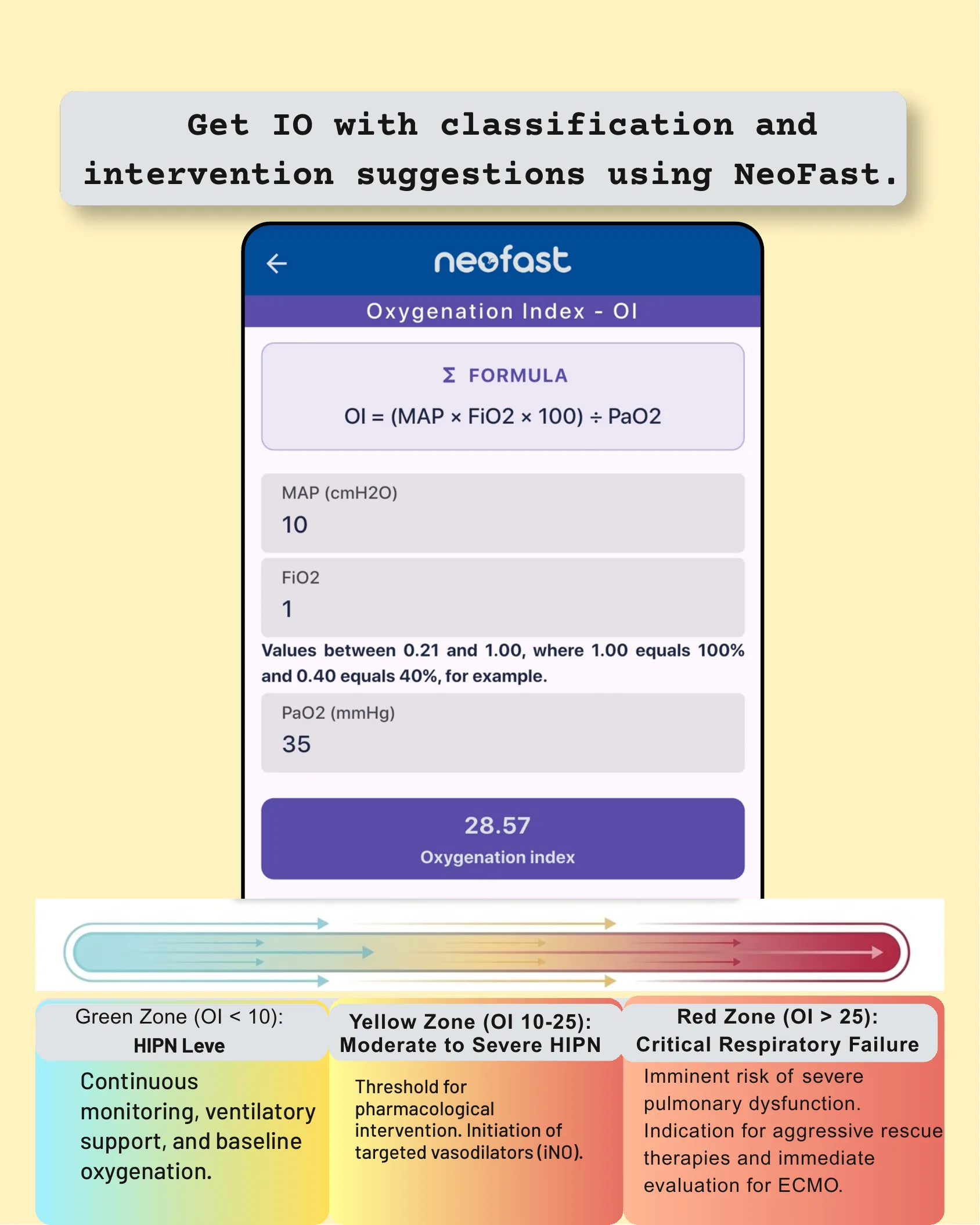
Get OI with classification and intervention suggestions using NeoFast
Σ Formula
OI = (MAP × FiO2 × 100) ÷ PaO2
- MAP (cmH2O): 10
- FiO2: 1 (Values between 0.21 and 1.00, where 1.00 equals 100% and 0.40 equals 40%, for example)
- PaO2 (mmHg): 35
28.57 Oxygenation index
| Zone | Classification | Intervention |
|---|---|---|
| Green Zone (OI < 10) | HIPN Leve | Continuous monitoring, ventilatory support, and baseline oxygenation. |
| Yellow Zone (OI 10-25) | Moderate to Severe HIPN | Threshold for pharmacological intervention. Initiation of targeted vasodilators (iNO). |
| Red Zone (OI > 25) | Critical Respiratory Failure | Imminent risk of severe pulmonary dysfunction. Indication for aggressive rescue therapies and immediate evaluation for ECMO. |
The Strategic Role of Biomarkers
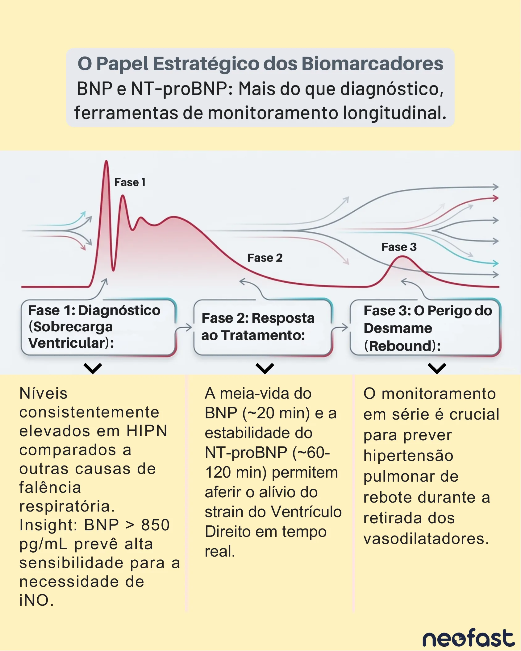
BNP and NT-proBNP: More than just diagnosis, tools for longitudinal monitoring
- Phase 1: Diagnosis (Ventricular Overload) – Consistently elevated levels in HIPN compared to other causes of respiratory failure. Insight: BNP > 850 pg/mL predicts high sensitivity to the need for iNO.
- Phase 2: Response to Treatment – The half-life of BNP (~20 min) and the stability of NT-proBNP (~60-120 min) allow for real-time assessment of right ventricular strain relief.
- Phase 3: The Danger of Weaning (Rebound) – Serial monitoring is crucial for predicting rebound pulmonary hypertension during vasodilator withdrawal.
Fundamentals of Management
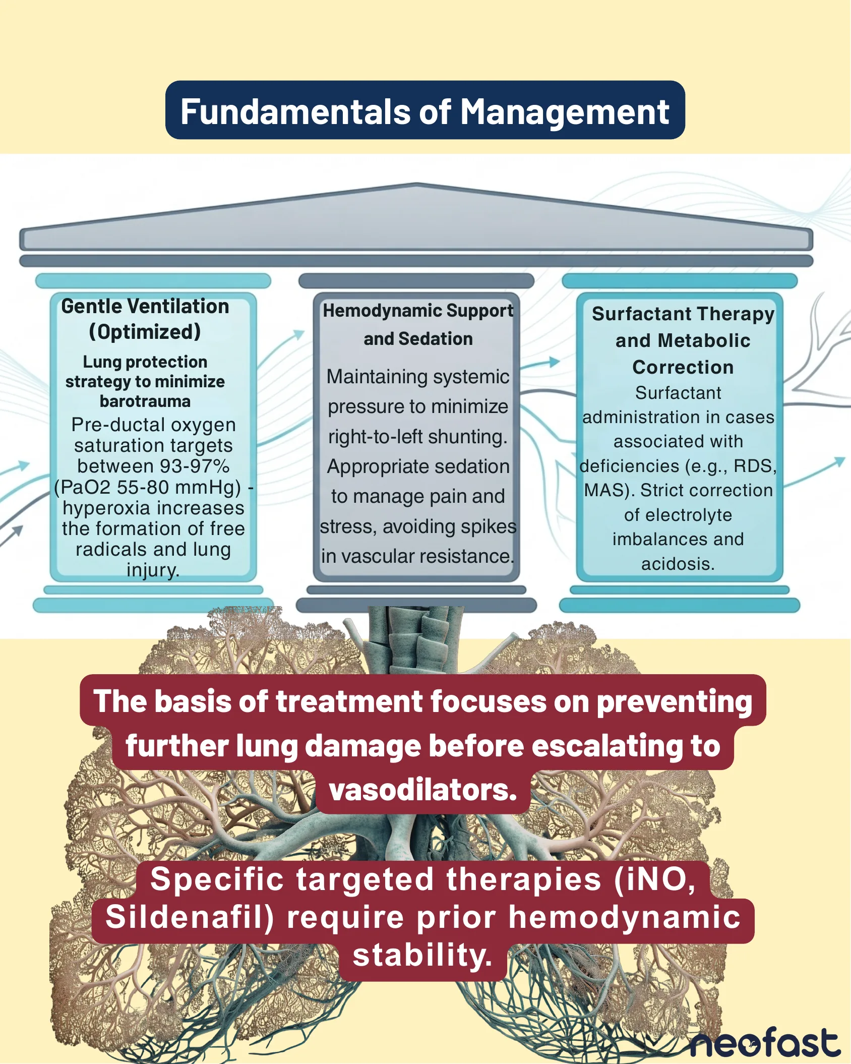
Fundamentals of Management
- Gentle Ventilation (Optimized): Lung protection strategy to minimize barotrauma. Pre-ductal oxygen saturation targets between 93-97% (PaO2 55-80 mmHg) – hyperoxia increases the formation of free radicals and lung injury.
- Hemodynamic Support and Sedation: Maintaining systemic pressure to minimize right-to-left shunting. Appropriate sedation to manage pain and stress, avoiding spikes in vascular resistance.
- Surfactant Therapy and Metabolic Correction: Surfactant administration in cases associated with deficiencies (e.g., RDS, MAS). Strict correction of electrolyte imbalances and acidosis.
The basis of treatment focuses on preventing further lung damage before escalating to vasodilators.
Specific targeted therapies (iNO, Sildenafil) require prior hemodynamic stability.
First-Line Therapy: Inhaled Nitric Oxide (iNO)
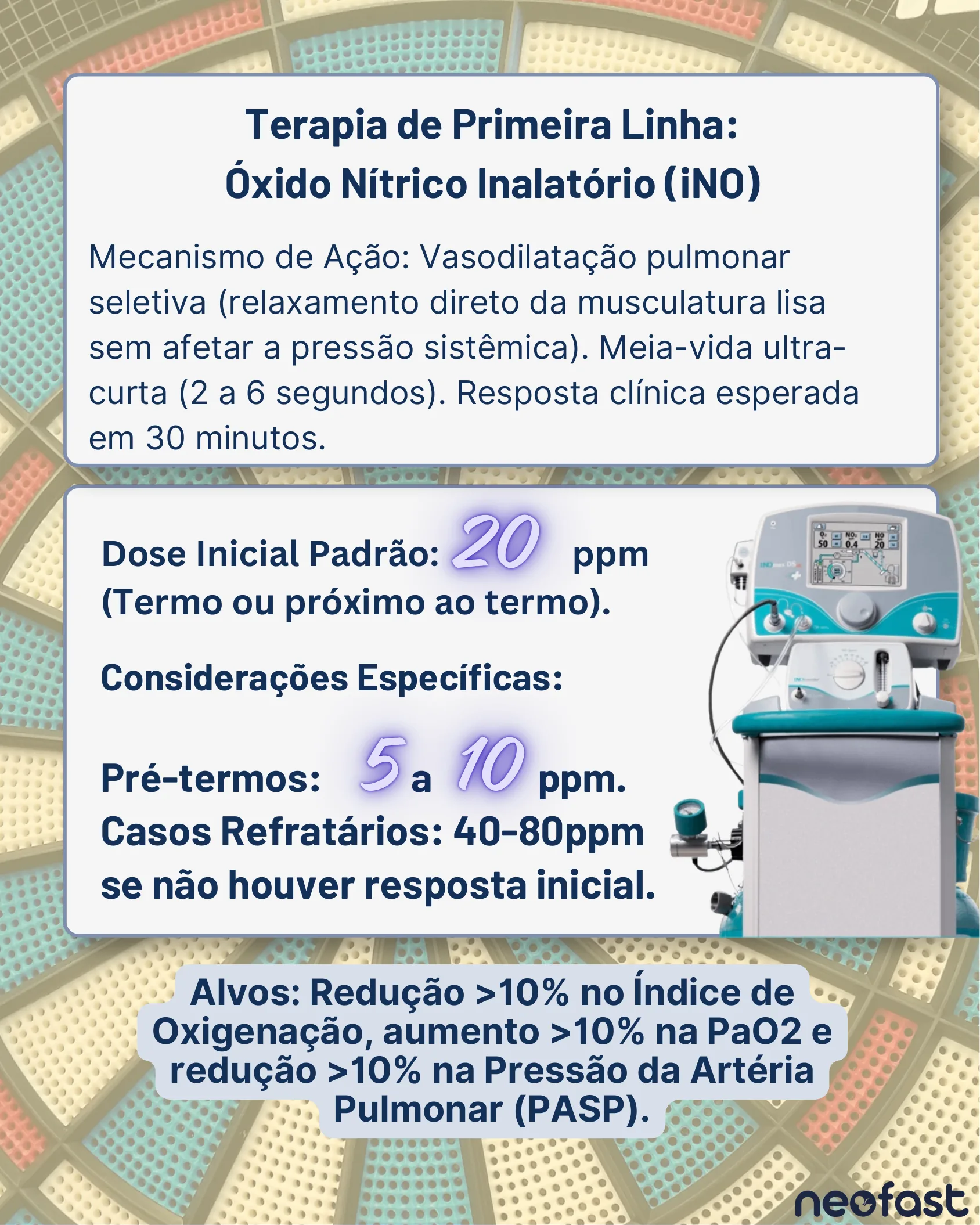
First-Line Therapy: Inhaled Nitric Oxide (iNO)
Mechanism of Action: Selective pulmonary vasodilation (direct relaxation of smooth muscle without affecting systemic pressure). Ultra-short half-life (2 to 6 seconds). Expected clinical response within 30 minutes.
- Standard Initial Dose: 20 ppm (Term or near term)
- Specific Considerations:
- Preterms: 5 to 10 ppm
- Refractory cases: 40-80 ppm if there is no initial response
Targets: Reduction >10% in Oxygenation Index, increase >10% in PaO2, and reduction >10% in Pulmonary Artery Pressure (PASP).
Targeted Vasodilators I: Sildenafil – Loading Dose
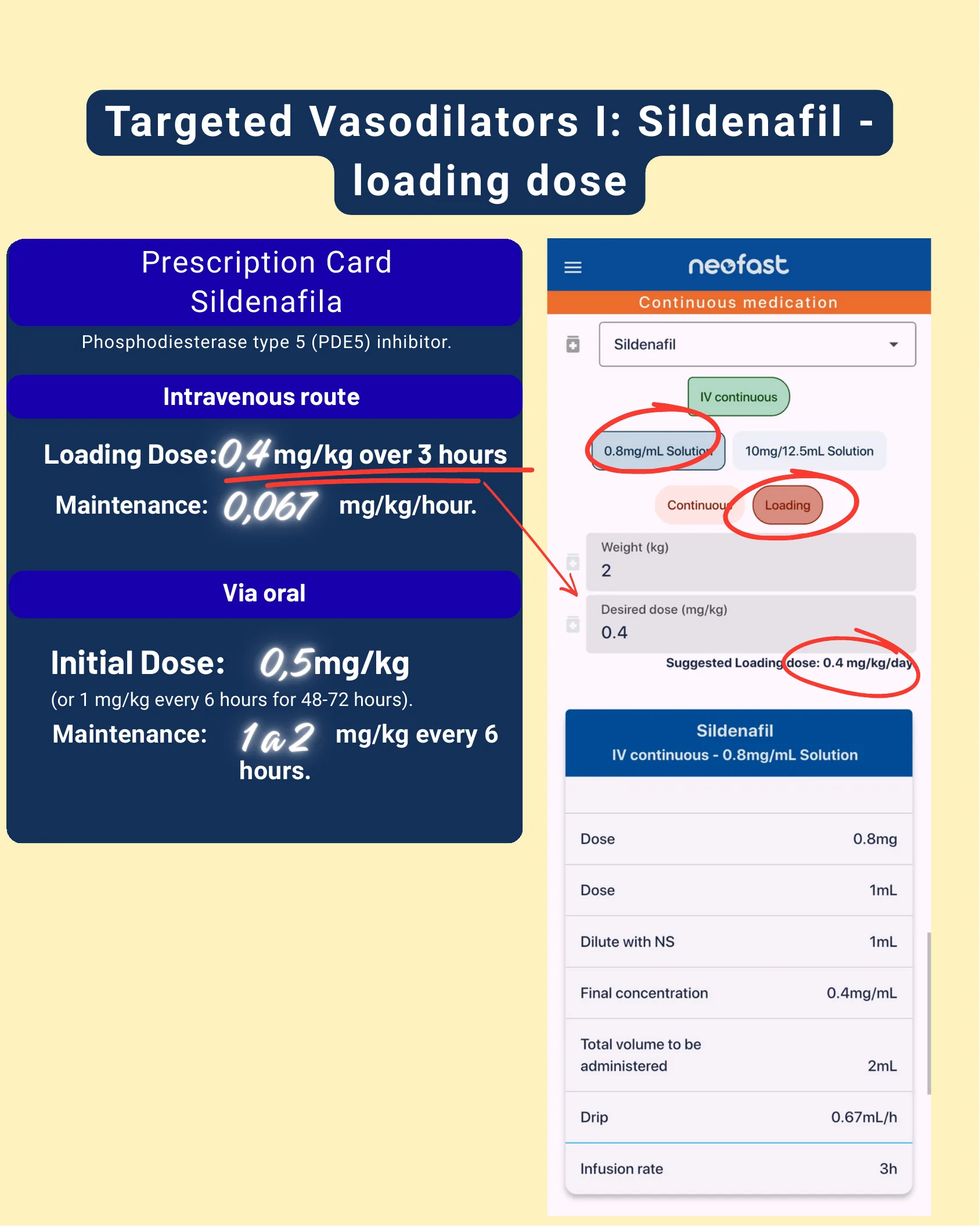
Targeted Vasodilators I: Sildenafil – loading dose
Prescription Card Sildenafil
Phosphodiesterase type 5 (PDE5) inhibitor.
Intravenous route
- Loading Dose: 0.4 mg/kg over 3 hours
- Maintenance: 0.067 mg/kg/hour
Via oral
- Initial Dose: 0.5 mg/kg (or 1 mg/kg every 6 hours for 48-72 hours)
- Maintenance: 1 to 2 mg/kg every 6 hours
NeoFast App Example (Loading)
| Field | Value |
|---|---|
| Medication | Sildenafil |
| Route | IV continuous – 0.8mg/mL Solution |
| Weight (kg) | 2 |
| Desired dose (mg/kg) | 0.4 |
| Suggested Loading dose | 0.4 mg/kg/day |
| Dose | 0.8mg |
| Dose (volume) | 1mL |
| Dilute with NS | 1mL |
| Final concentration | 0.4mg/mL |
| Total volume to be administered | 2mL |
| Drip | 0.67mL/h |
| Infusion rate | 3h |
Targeted Vasodilators I: Continuous Sildenafil
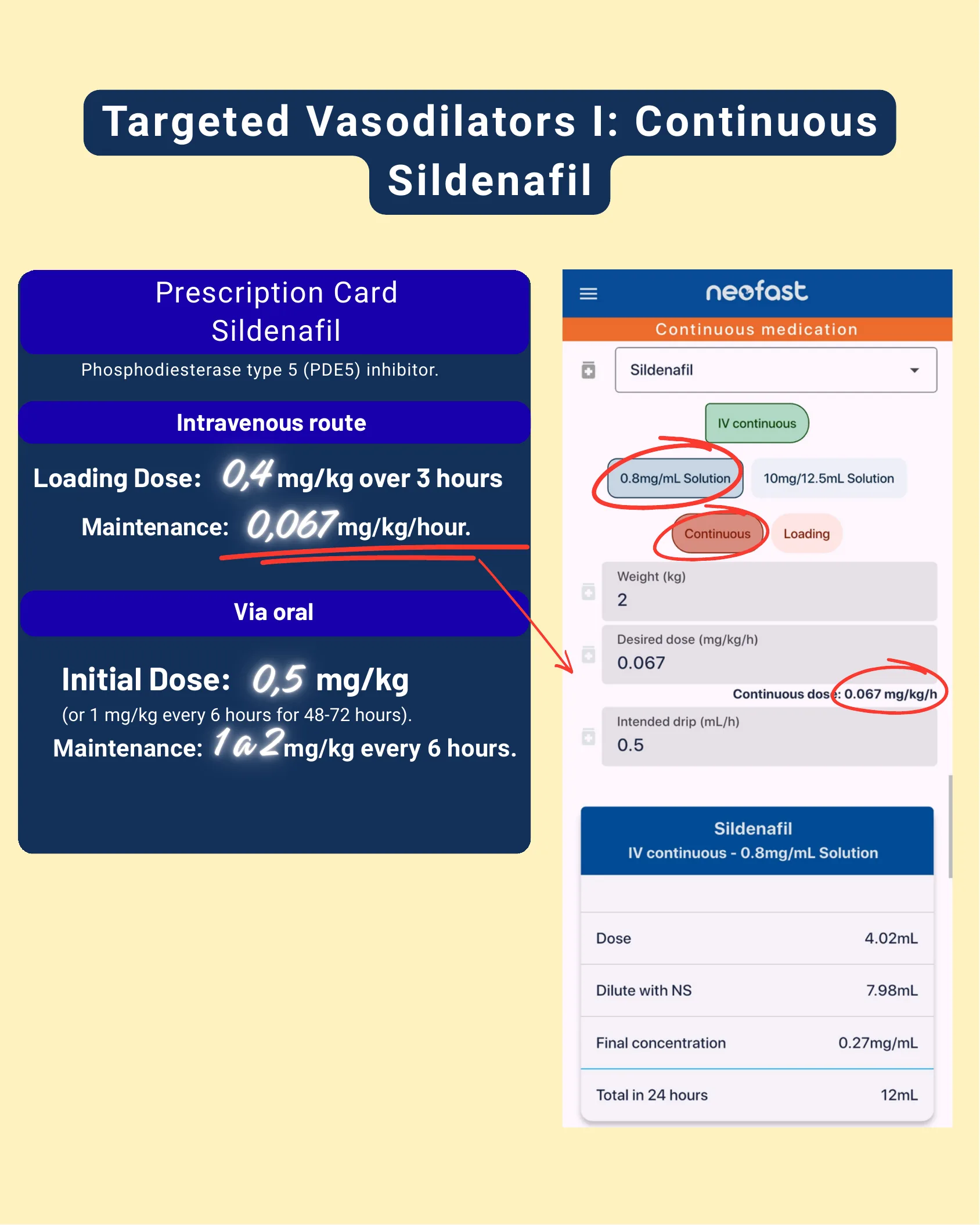
Targeted Vasodilators I: Continuous Sildenafil
Prescription Card Sildenafil
Phosphodiesterase type 5 (PDE5) inhibitor.
Intravenous route
- Loading Dose: 0.4 mg/kg over 3 hours
- Maintenance: 0.067 mg/kg/hour
Via oral
- Initial Dose: 0.5 mg/kg (or 1 mg/kg every 6 hours for 48-72 hours)
- Maintenance: 1 to 2 mg/kg every 6 hours
NeoFast App Example (Continuous)
| Field | Value |
|---|---|
| Medication | Sildenafil |
| Route | IV continuous – 0.8mg/mL Solution |
| Weight (kg) | 2 |
| Desired dose (mg/kg/h) | 0.067 |
| Continuous dose | 0.067 mg/kg/h |
| Intended drip (mL/h) | 0.5 |
| Dose | 4.02mL |
| Dilute with NS | 7.98mL |
| Final concentration | 0.27mg/mL |
| Total in 24 hours | 12mL |
Targeted Vasodilators I: Continuous Milrinone
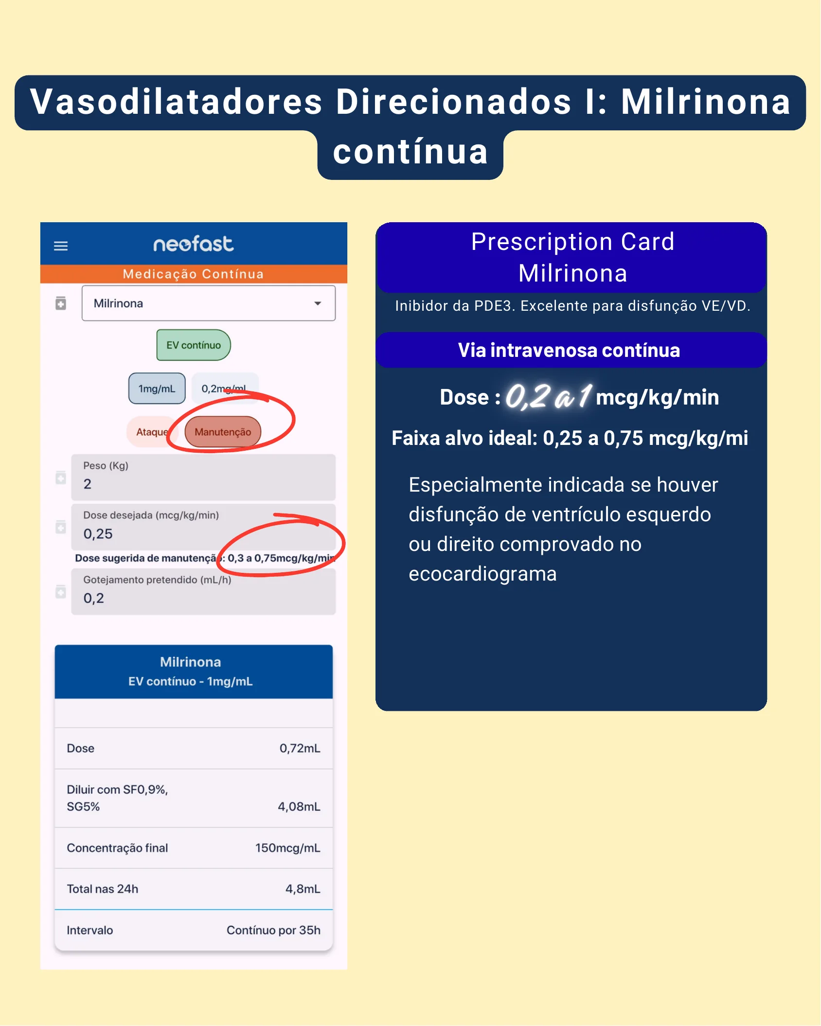
Targeted Vasodilators I: Continuous Milrinone
Prescription Card Milrinone
PDE3 inhibitor. Excellent for left ventricular/right ventricular dysfunction.
Continuous intravenous administration
- Dose: 0.2 to 1 mcg/kg/min
- Ideal target range: 0.25 to 0.75 mcg/kg/min
It is especially indicated if left or right ventricular dysfunction is confirmed by echocardiography.
NeoFast App Example (Maintenance)
| Field | Value |
|---|---|
| Medication | Milrinone |
| Route | IV continuous – 1mg/mL |
| Weight (kg) | 2 |
| Desired dose (mcg/kg/min) | 0.25 |
| Suggested maintenance dose | 0.3 to 0.75 mcg/kg/min |
| Intended drip (mL/h) | 0.2 |
| Dose | 0.72mL |
| Dilute with NS, D5W | 4.08mL |
| Final concentration | 150mcg/mL |
| Total in 24 hours | 4.8mL |
| Interval | Continuous by 35h |
Targeted Vasodilators II: Bosentan, Prostanoids, and Hemodynamic Support
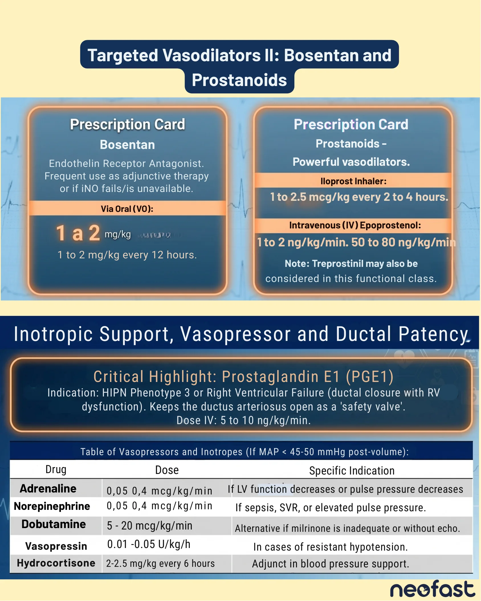
Targeted Vasodilators II: Bosentan and Prostanoids
Prescription Card Bosentan
Endothelin Receptor Antagonist. Frequent use as adjunctive therapy or if iNO fails/is unavailable.
Via Oral (VO): 1 to 2 mg/kg every 12 hours.
Prescription Card Prostanoids – Powerful vasodilators
- Iloprost Inhaler: 1 to 2.5 mcg/kg every 2 to 4 hours.
- Intravenous (IV) Epoprostenol: 1 to 2 ng/kg/min. 50 to 80 ng/kg/min
Note: Treprostinil may also be considered in this functional class.
Inotropic Support, Vasopressor and Ductal Patency
Critical Highlight: Prostaglandin E1 (PGE1)
Indication: HIPN Phenotype 3 or Right Ventricular Failure (ductal closure with RV dysfunction). Keeps the ductus arteriosus open as a ‘safety valve’. Dose IV: 5 to 10 ng/kg/min.
Table of Vasopressors and Inotropes (If MAP < 45-50 mmHg post-volume)
| Drug | Dose | Specific Indication |
|---|---|---|
| Adrenaline | 0.05-0.4 mcg/kg/min | If LV function decreases or pulse pressure decreases |
| Norepinephrine | 0.05-0.4 mcg/kg/min | If sepsis, SVR, or elevated pulse pressure. |
| Dobutamine | 5-20 mcg/kg/min | Alternative if milrinone is inadequate or without echo. |
| Vasopressin | 0.01-0.05 U/kg/h | In cases of resistant hypotension. |
| Hydrocortisone | 2-2.5 mg/kg every 6 hours | Adjunct in blood pressure support. |
PGE1 Continuous Medication Calculator
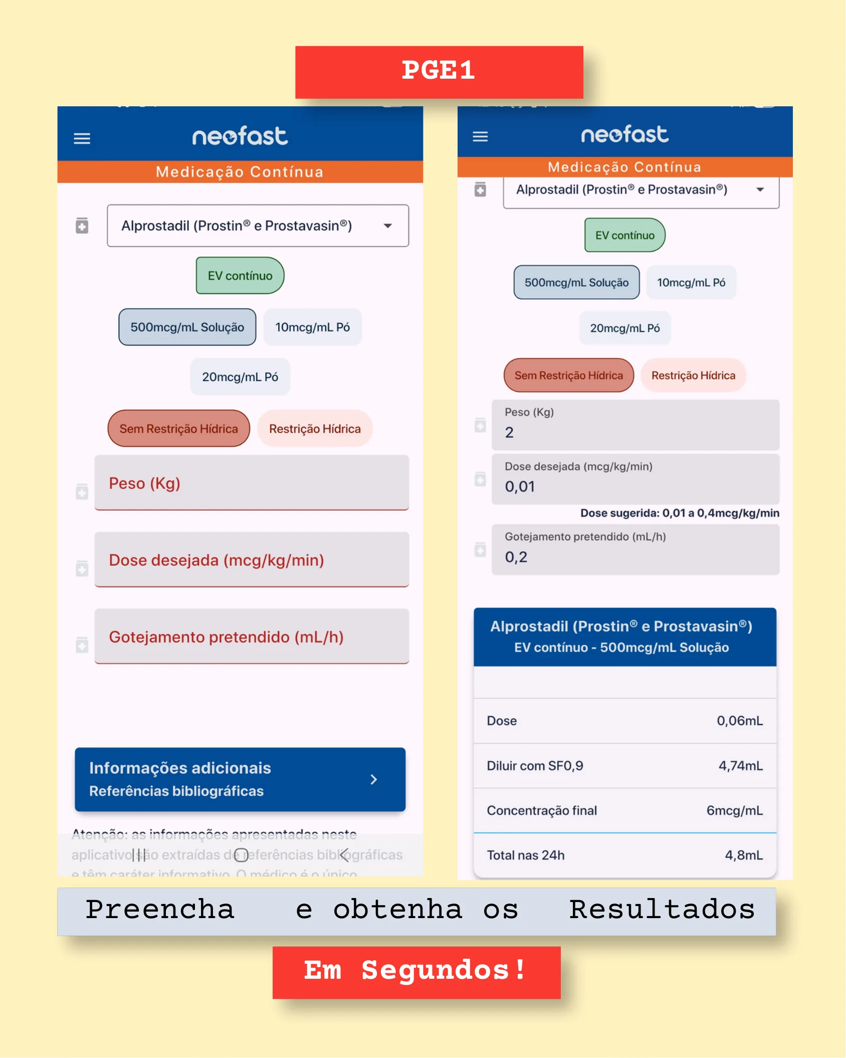
PGE1
Using the NeoFast app for Alprostadil (Prostin® and Prostavasin®) continuous medication:
- Select drug: Alprostadil (Prostin® and Prostavasin®)
- Route: IV continuous
- Concentration options: 500mcg/mL Solution, 10mcg/mL Powder, 20mcg/mL Powder
- Fluid option: No Fluid Restriction / Fluid restriction
- Input fields: Weight (kg), Desired dose (mcg/kg/min), Intended drip (mL/h)
- Additional information / Bibliographical references available
Example calculation:
- Weight (kg): 2
- Desired dose (mcg/kg/min): 0.01 (Suggested dose: 0.01 to 0.4 mcg/kg/min)
- Intended drip (mL/h): 0.2
Result: Alprostadil (Prostin® and Prostavasin®) – IV continuous – 500mcg/mL Solution
| Parameter | Value |
|---|---|
| Dose | 0.06mL |
| Dilute with NS | 4.74mL |
| Final concentration | 6mcg/mL |
| Total in 24 hours | 4.8mL |
Fill in → and obtain the → Results
In seconds!
Adrenaline and Noradrenaline
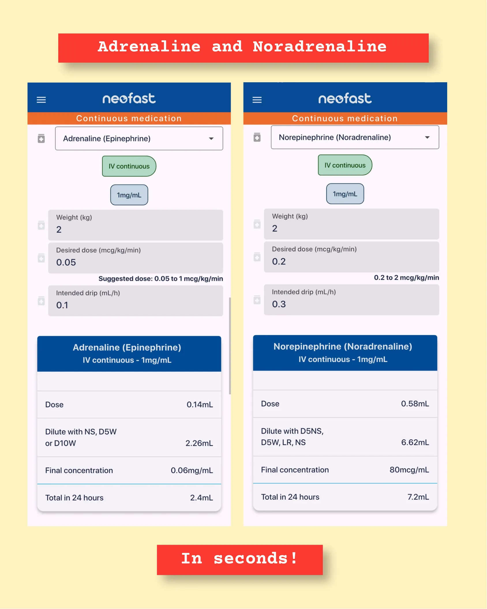
Adrenaline and Noradrenaline
Adrenaline (Epinephrine)
- Route: IV continuous
- Concentration: 1mg/mL
- Weight (kg): 2
- Desired dose (mcg/kg/min): 0.05 (Suggested dose: 0.05 to 1 mcg/kg/min)
- Intended drip (mL/h): 0.1
| Parameter | Value |
|---|---|
| Dose | 0.14mL |
| Dilute with NS, D5W or D10W | 2.26mL |
| Final concentration | 0.06mg/mL |
| Total in 24 hours | 2.4mL |
Norepinephrine (Noradrenaline)
- Route: IV continuous
- Concentration: 1mg/mL
- Weight (kg): 2
- Desired dose (mcg/kg/min): 0.2 (0.2 to 2 mcg/kg/min)
- Intended drip (mL/h): 0.3
| Parameter | Value |
|---|---|
| Dose | 0.58mL |
| Dilute with D5NS, D5W, LR, NS | 6.62mL |
| Final concentration | 80mcg/mL |
| Total in 24 hours | 7.2mL |
In seconds!
Dobutamine and Vasopressin
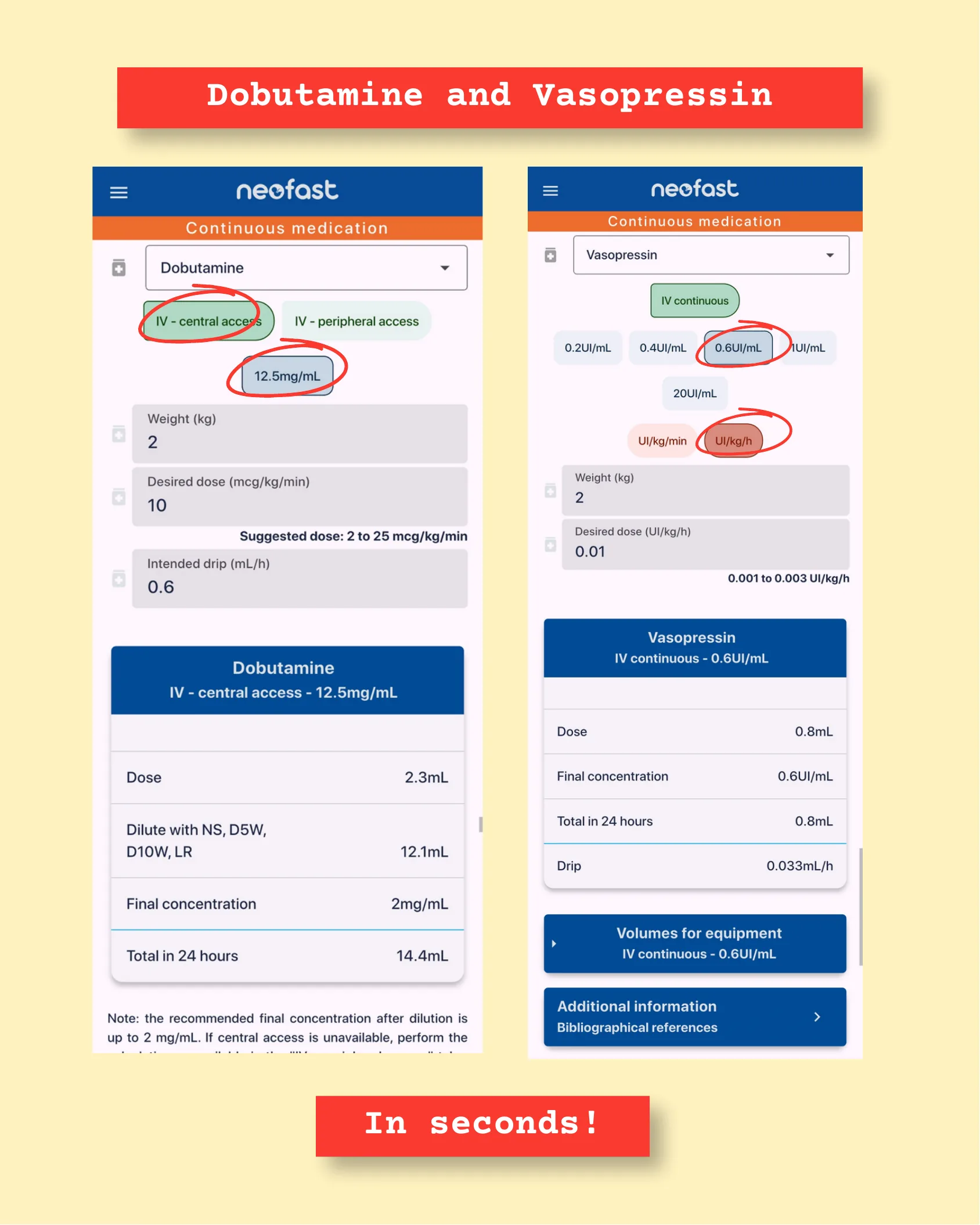
Dobutamine and Vasopressin
Dobutamine
- Route: IV – central access (circled/selected over IV – peripheral access)
- Concentration: 12.5mg/mL (circled/selected)
- Weight (kg): 2
- Desired dose (mcg/kg/min): 10 (Suggested dose: 2 to 25 mcg/kg/min)
- Intended drip (mL/h): 0.6
| Parameter | Value |
|---|---|
| Dose | 2.3mL |
| Dilute with NS, D5W, D10W, LR | 12.1mL |
| Final concentration | 2mg/mL |
| Total in 24 hours | 14.4mL |
Note: the recommended final concentration after dilution is up to 2 mg/mL. If central access is unavailable, perform the calculation available in the
Frequently asked questions
What is the formula for calculating the Oxygenation Index (OI) in PPHN?
OI = (MAP × FiO2 × 100) ÷ PaO2, where MAP is in cmH2O, FiO2 ranges from 0.21 to 1.00, and PaO2 is in mmHg. An OI less than 10 is the Green Zone (mild PPHN, monitoring), OI 10-25 is the Yellow Zone (moderate-severe, threshold for iNO), and OI greater than 25 is the Red Zone (critical respiratory failure, indication for aggressive rescue therapy and ECMO evaluation).
What is the dosing for inhaled nitric oxide (iNO) in PPHN?
Standard initial dose is 20 ppm for term or near-term infants. Preterm infants receive 5 to 10 ppm. Refractory cases may require 40-80 ppm if there is no initial response. Targets include a reduction greater than 10% in Oxygenation Index, an increase greater than 10% in PaO2, and a reduction greater than 10% in Pulmonary Artery Pressure (PASP).
What is the IV sildenafil loading and maintenance dose for PPHN?
Loading dose is 0.4 mg/kg IV over 3 hours, followed by a maintenance dose of 0.067 mg/kg/hour. Oral dosing alternatives include an initial dose of 0.5 mg/kg (or 1 mg/kg every 6 hours for 48-72 hours) with maintenance of 1 to 2 mg/kg every 6 hours.
When should milrinone be used in PPHN, and at what dose?
Milrinone, a PDE3 inhibitor, is especially indicated when left or right ventricular dysfunction is confirmed by echocardiography. Continuous IV dose ranges from 0.2 to 1 mcg/kg/min, with an ideal target range of 0.25 to 0.75 mcg/kg/min.
What is the role of Prostaglandin E1 (PGE1) in PPHN management?
PGE1 is indicated in PPHN Phenotype 3 or right ventricular failure to keep the ductus arteriosus open as a ‘safety valve’ when ductal closure occurs with RV dysfunction. The IV dose is 5 to 10 ng/kg/min.