Key takeaways
- Neonatal shock occurs when oxygen delivery (DO₂ = CO × CaO₂) fails to meet tissue metabolic demand (VO₂), triggering anaerobic metabolism and lactic acidosis.
- Because neonatal stroke volume is small and fixed, cardiac output is heart-rate dependent, but tachycardia above 180 bpm shortens diastole, reduces ventricular filling and causes subendocardial ischemia (blunted Bowditch effect).
- Capillary refill time ≥3 seconds has only 55% sensitivity and 80% specificity, meaning 7 in 10 neonates with low SVC flow have a normal CRT, so normotension does not equal adequate perfusion.
- Fluid boluses should be 10 mL/kg of 0.9% saline, infused over 15-30 minutes in term infants and 30-60 minutes in preterm infants (maximum 10-20 mL/kg total), and reserved for confirmed hypovolemia on TnECHO.
- In refractory shock despite fluids and escalating vasoactive doses, hydrocortisone 1 mg/kg every 6-8 h is indicated for relative adrenal insufficiency of prematurity.
References
- Nouraeyan N, Wong K, Kharrat A, Fraga MV, Altit G. A comprehensive review of the approach to ultrasonography in neonatal shock: targeted echocardiography and POCUS. Children. 2026;13:1268.
- Davis AL, Carcillo JA, Aneja RK, et al. ACCM clinical practice parameters for hemodynamic support of pediatric and neonatal septic shock. Crit Care Med. 2017;45(6):1061-93.
- McNamara PJ, Jain A, El-Khuffash A, et al. Guidelines and recommendations for targeted neonatal echocardiography and cardiac POCUS in the NICU: update from the ASE. J Am Soc Echocardiogr. 2024;37:171-215.
- Weiss SL, Peters MJ, Alhazzani W, et al. Surviving Sepsis Campaign international guidelines for the management of septic shock in children. Pediatr Crit Care Med. 2020;21(2):e52-e106.
- Giesinger RE, McNamara PJ. Hemodynamic instability in the critically ill neonate: an approach based on disease pathophysiology. Semin Perinatol. 2016;40(3):174-88.

NEONATAL SHOCK: PATHOPHYSIOLOGY AND MANAGEMENT
For Neonatologists
A complete, up-to-date guide to early recognition, advanced pathophysiology, differentiation of hemodynamic phenotypes and targeted management in the NICU.
The Concept of Shock and the DO₂/VO₂ Balance
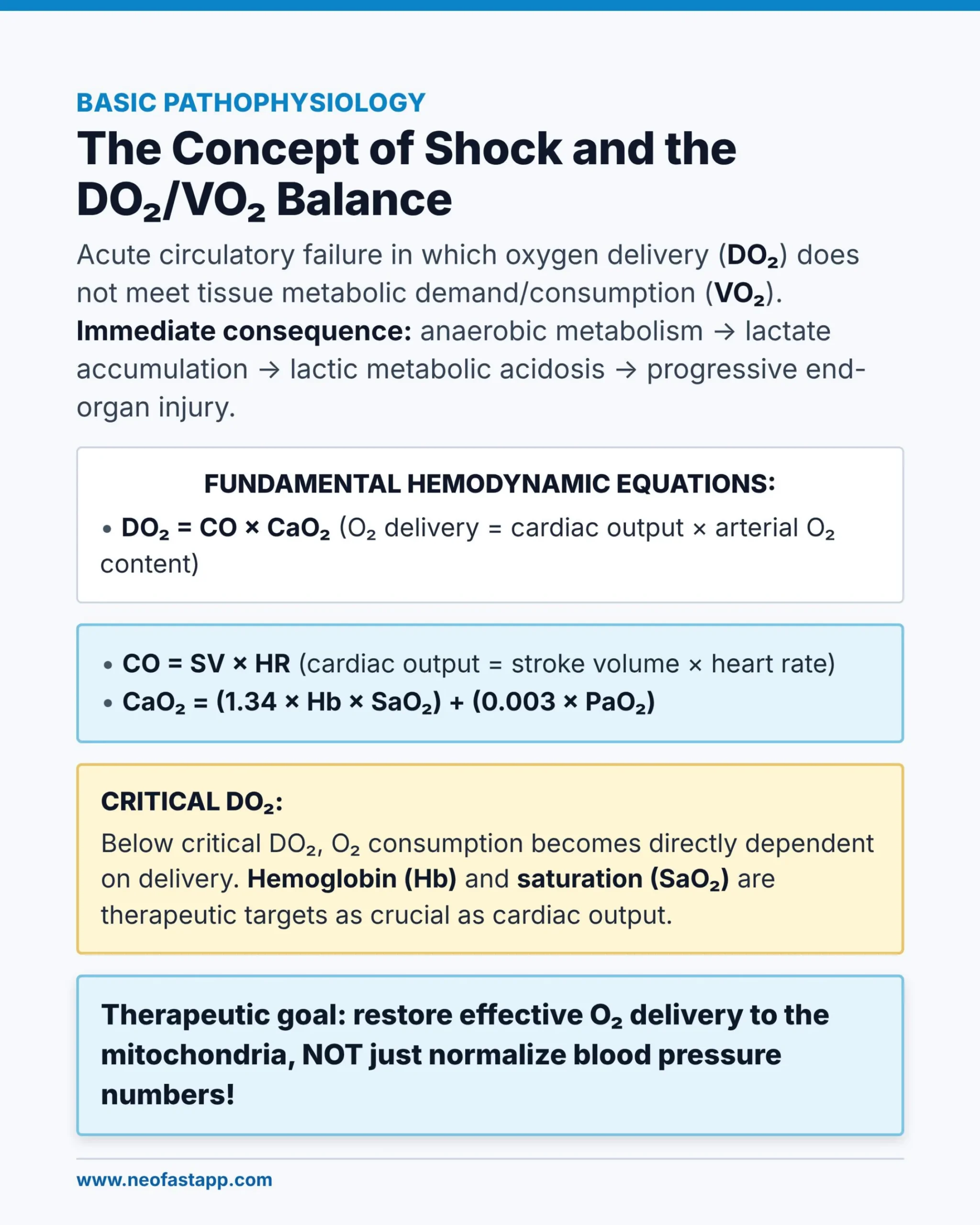
BASIC PATHOPHYSIOLOGY
The Concept of Shock and the DO₂/VO₂ Balance
Acute circulatory failure in which oxygen delivery (DO₂) does not meet tissue metabolic demand/consumption (VO₂). Immediate consequence: anaerobic metabolism → lactate accumulation → lactic metabolic acidosis → progressive end-organ injury.
FUNDAMENTAL HEMODYNAMIC EQUATIONS:
- DO₂ = CO × CaO₂ (O₂ delivery = cardiac output × arterial O₂ content)
- CO = SV × HR (cardiac output = stroke volume × heart rate)
- CaO₂ = (1.34 × Hb × SaO₂) + (0.003 × PaO₂)
CRITICAL DO₂:
Below critical DO₂, O₂ consumption becomes directly dependent on delivery. Hemoglobin (Hb) and saturation (SaO₂) are therapeutic targets as crucial as cardiac output.
Therapeutic goal: restore effective O₂ delivery to the mitochondria, NOT just normalize blood pressure numbers!
What Makes the Neonatal Heart Different
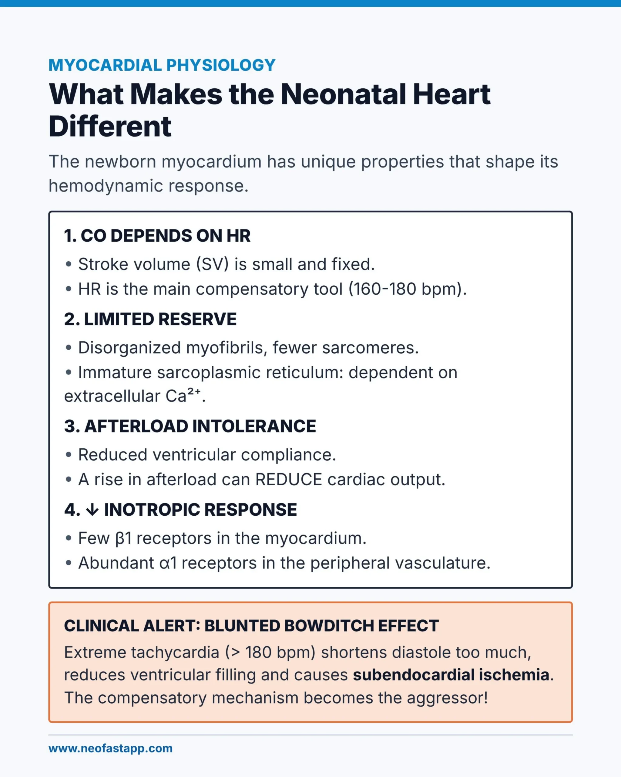
MYOCARDIAL PHYSIOLOGY
What Makes the Neonatal Heart Different
The newborn myocardium has unique properties that shape its hemodynamic response.
1. CO DEPENDS ON HR
- Stroke volume (SV) is small and fixed.
- HR is the main compensatory tool (160-180 bpm).
2. LIMITED RESERVE
- Disorganized myofibrils, fewer sarcomeres.
- Immature sarcoplasmic reticulum: dependent on extracellular Ca²⁺.
3. AFTERLOAD INTOLERANCE
- Reduced ventricular compliance.
- A rise in afterload can REDUCE cardiac output.
4. ↓ INOTROPIC RESPONSE
- Few β1 receptors in the myocardium.
- Abundant α1 receptors in the peripheral vasculature.
CLINICAL ALERT: BLUNTED BOWDITCH EFFECT
Extreme tachycardia (> 180 bpm) shortens diastole too much, reduces ventricular filling and causes subendocardial ischemia. The compensatory mechanism becomes the aggressor!
Circulatory Transition and Shunts
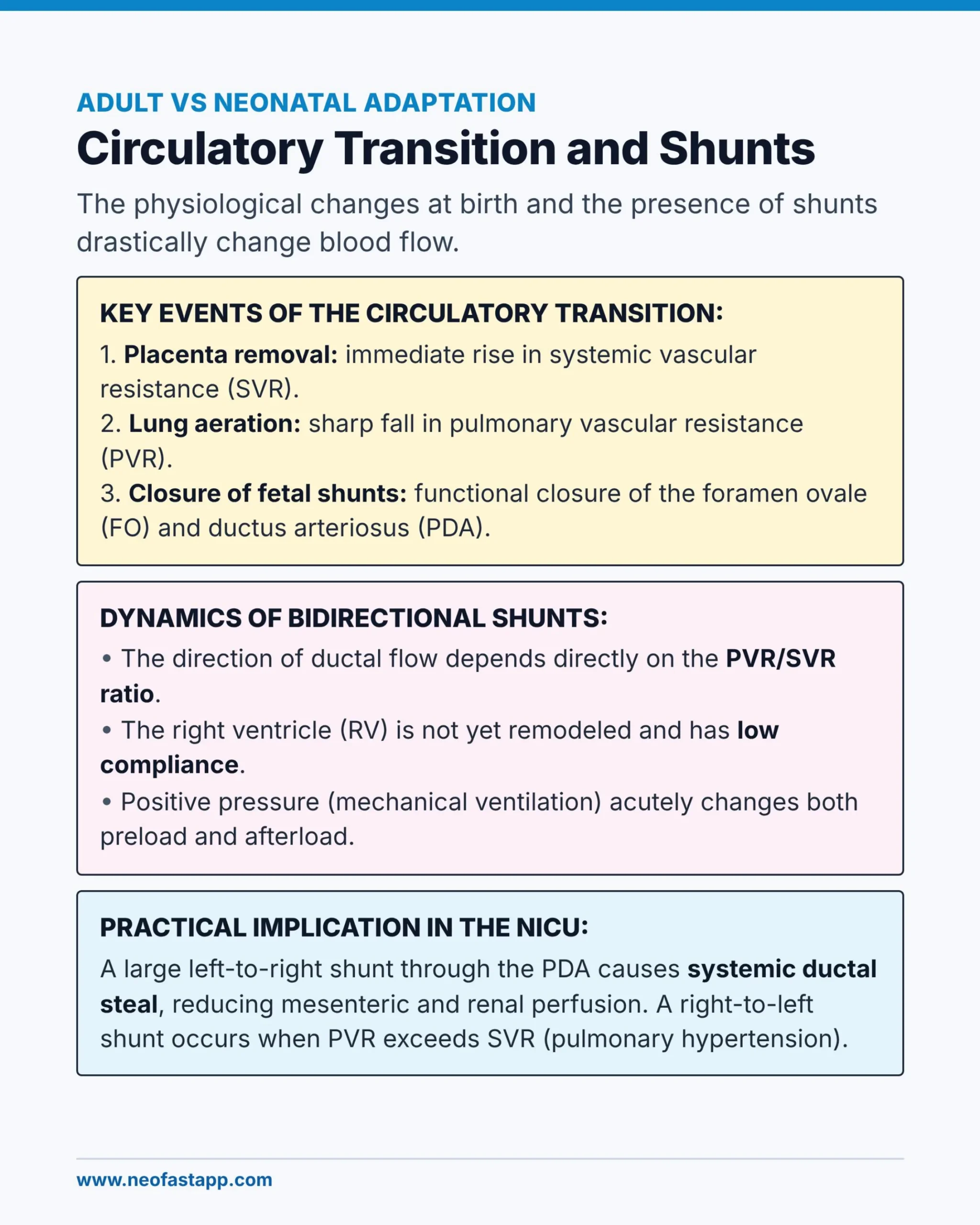
ADULT VS NEONATAL ADAPTATION
Circulatory Transition and Shunts
The physiological changes at birth and the presence of shunts drastically change blood flow.
KEY EVENTS OF THE CIRCULATORY TRANSITION:
- Placenta removal: immediate rise in systemic vascular resistance (SVR).
- Lung aeration: sharp fall in pulmonary vascular resistance (PVR).
- Closure of fetal shunts: functional closure of the foramen ovale (FO) and ductus arteriosus (PDA).
DYNAMICS OF BIDIRECTIONAL SHUNTS:
- The direction of ductal flow depends directly on the PVR/SVR ratio.
- The right ventricle (RV) is not yet remodeled and has low compliance.
- Positive pressure (mechanical ventilation) acutely changes both preload and afterload.
PRACTICAL IMPLICATION IN THE NICU:
A large left-to-right shunt through the PDA causes systemic ductal steal, reducing mesenteric and renal perfusion. A right-to-left shunt occurs when PVR exceeds SVR (pulmonary hypertension).
Etiological Phenotypes of Neonatal Shock
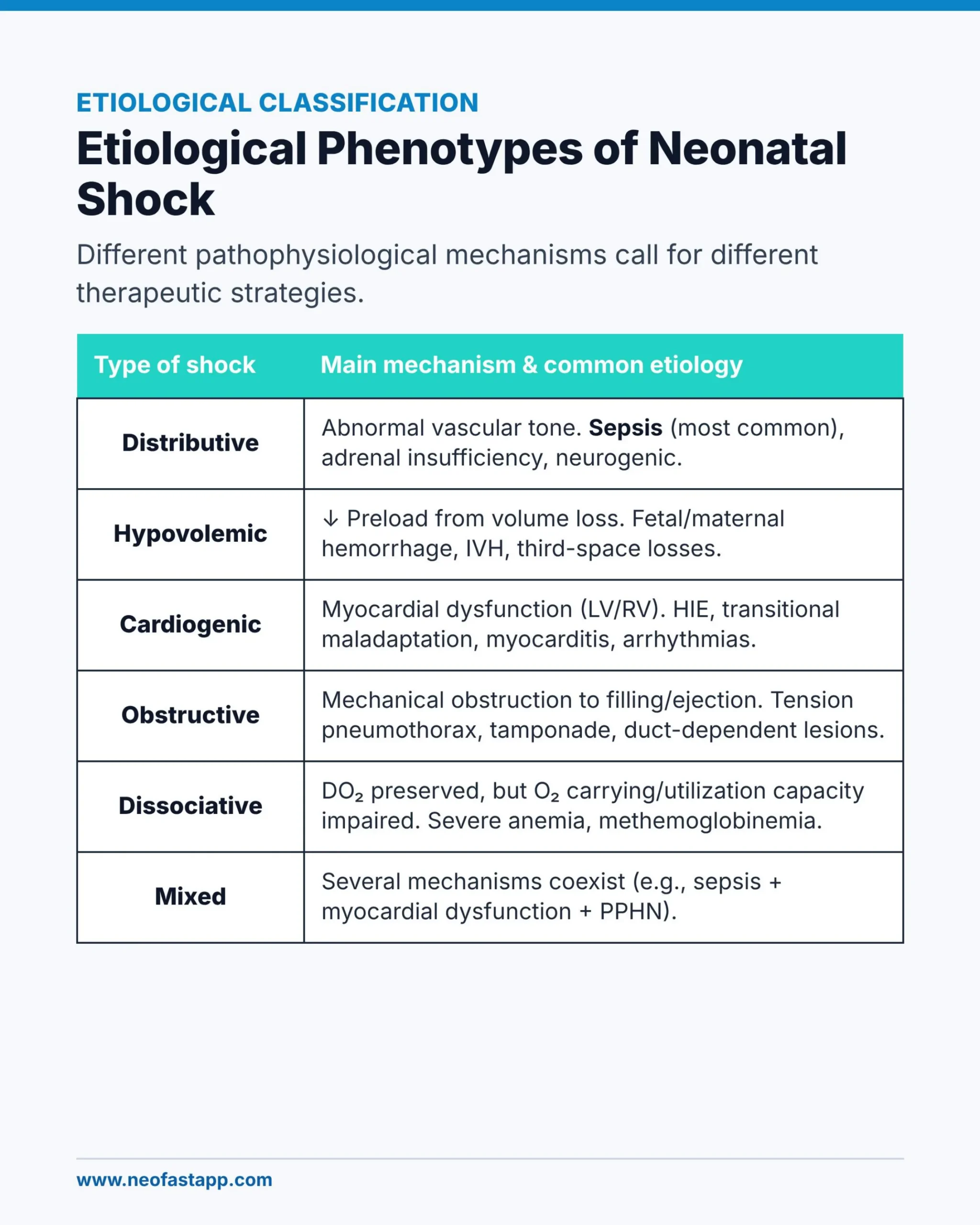
ETIOLOGICAL CLASSIFICATION
Etiological Phenotypes of Neonatal Shock
Different pathophysiological mechanisms call for different therapeutic strategies.
| Type of shock | Main mechanism & common etiology |
|---|---|
| Distributive | Abnormal vascular tone. Sepsis (most common), adrenal insufficiency, neurogenic. |
| Hypovolemic | ↓ Preload from volume loss. Fetal/maternal hemorrhage, IVH, third-space losses. |
| Cardiogenic | Myocardial dysfunction (LV/RV). HIE, transitional maladaptation, myocarditis, arrhythmias. |
| Obstructive | Mechanical obstruction to filling/ejection. Tension pneumothorax, tamponade, duct-dependent lesions. |
| Dissociative | DO₂ preserved, but O₂ carrying/utilization capacity impaired. Severe anemia, methemoglobinemia. |
| Mixed | Several mechanisms coexist (e.g., sepsis + myocardial dysfunction + PPHN). |
Septic Shock: Warm vs. Cold
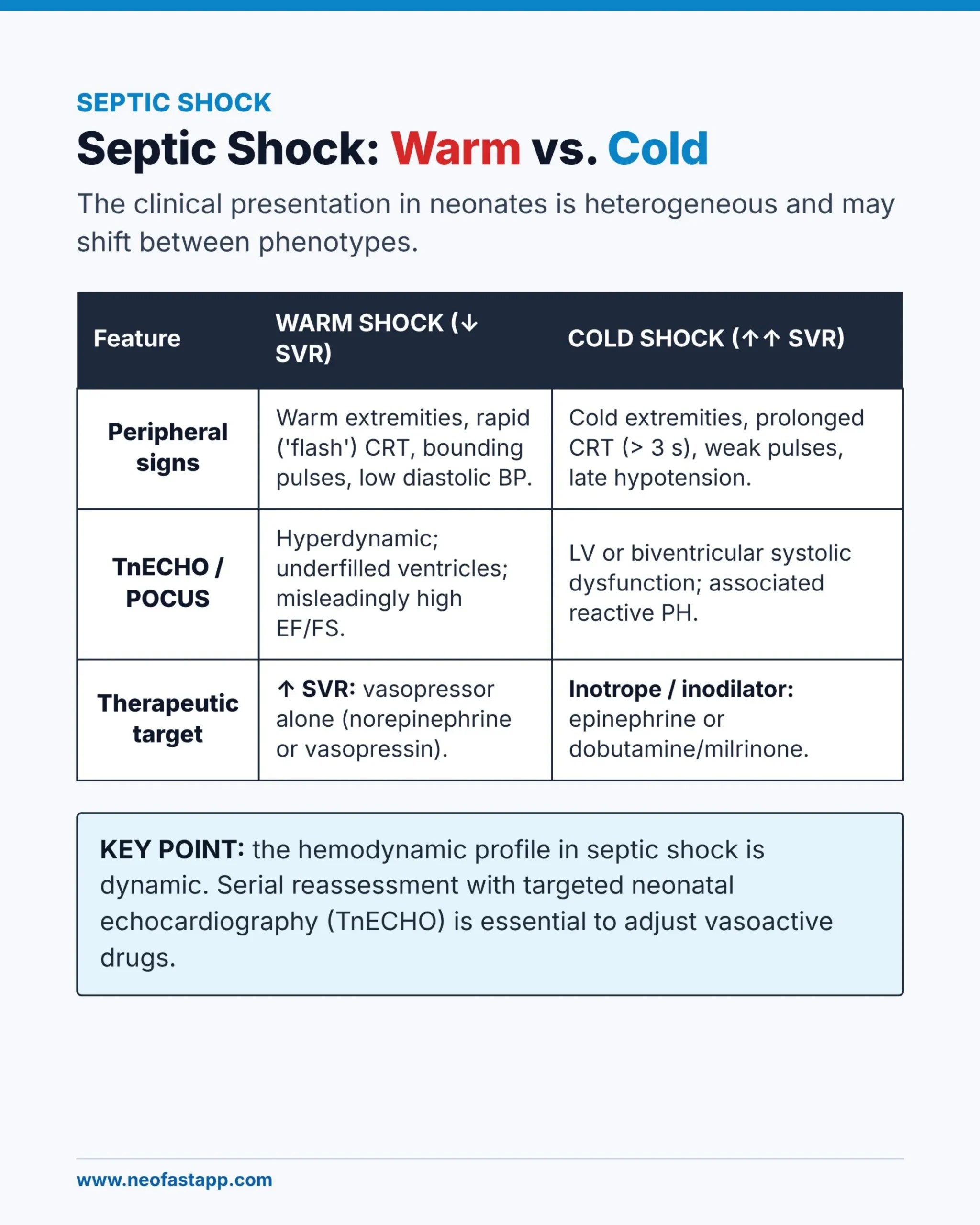
SEPTIC SHOCK
Septic Shock: Warm vs. Cold
The clinical presentation in neonates is heterogeneous and may shift between phenotypes.
| Feature | WARM SHOCK (↓ SVR) | COLD SHOCK (↑↑ SVR) |
|---|---|---|
| Peripheral signs | Warm extremities, rapid (‘flash’) CRT, bounding pulses, low diastolic BP. | Cold extremities, prolonged CRT (> 3 s), weak pulses, late hypotension. |
| TnECHO / POCUS | Hyperdynamic; underfilled ventricles; misleadingly high EF/FS. | LV or biventricular systolic dysfunction; associated reactive PH. |
| Therapeutic target | ↑ SVR: vasopressor alone (norepinephrine or vasopressin). | Inotrope / inodilator: epinephrine or dobutamine/milrinone. |
KEY POINT: the hemodynamic profile in septic shock is dynamic. Serial reassessment with targeted neonatal echocardiography (TnECHO) is essential to adjust vasoactive drugs.
Continuous Medication Calculator
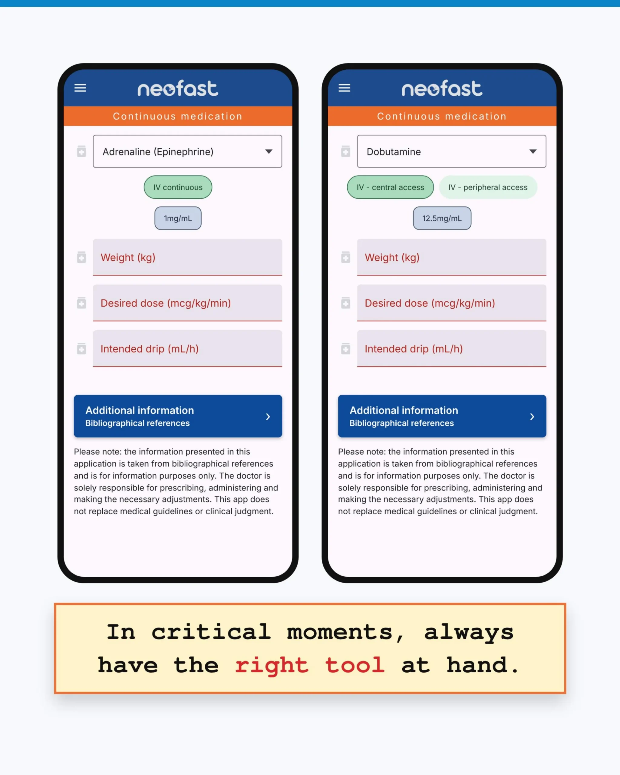
Continuous medication
Adrenaline (Epinephrine)
- IV continuous
- 1mg/mL
- Weight (kg)
- Desired dose (mcg/kg/min)
- Intended drip (mL/h)
Additional information – Bibliographical references
Please note: the information presented in this application is taken from bibliographical references and is for information purposes only. The doctor is solely responsible for prescribing, administering and making the necessary adjustments. This app does not replace medical guidelines or clinical judgment.
Dobutamine
- IV – central access
- IV – peripheral access
- 12.5mg/mL
- Weight (kg)
- Desired dose (mcg/kg/min)
- Intended drip (mL/h)
Additional information – Bibliographical references
Please note: the information presented in this application is taken from bibliographical references and is for information purposes only. The doctor is solely responsible for prescribing, administering and making the necessary adjustments. This app does not replace medical guidelines or clinical judgment.
In critical moments, always have the right tool at hand.
Continuous Medication Calculator – Milrinone
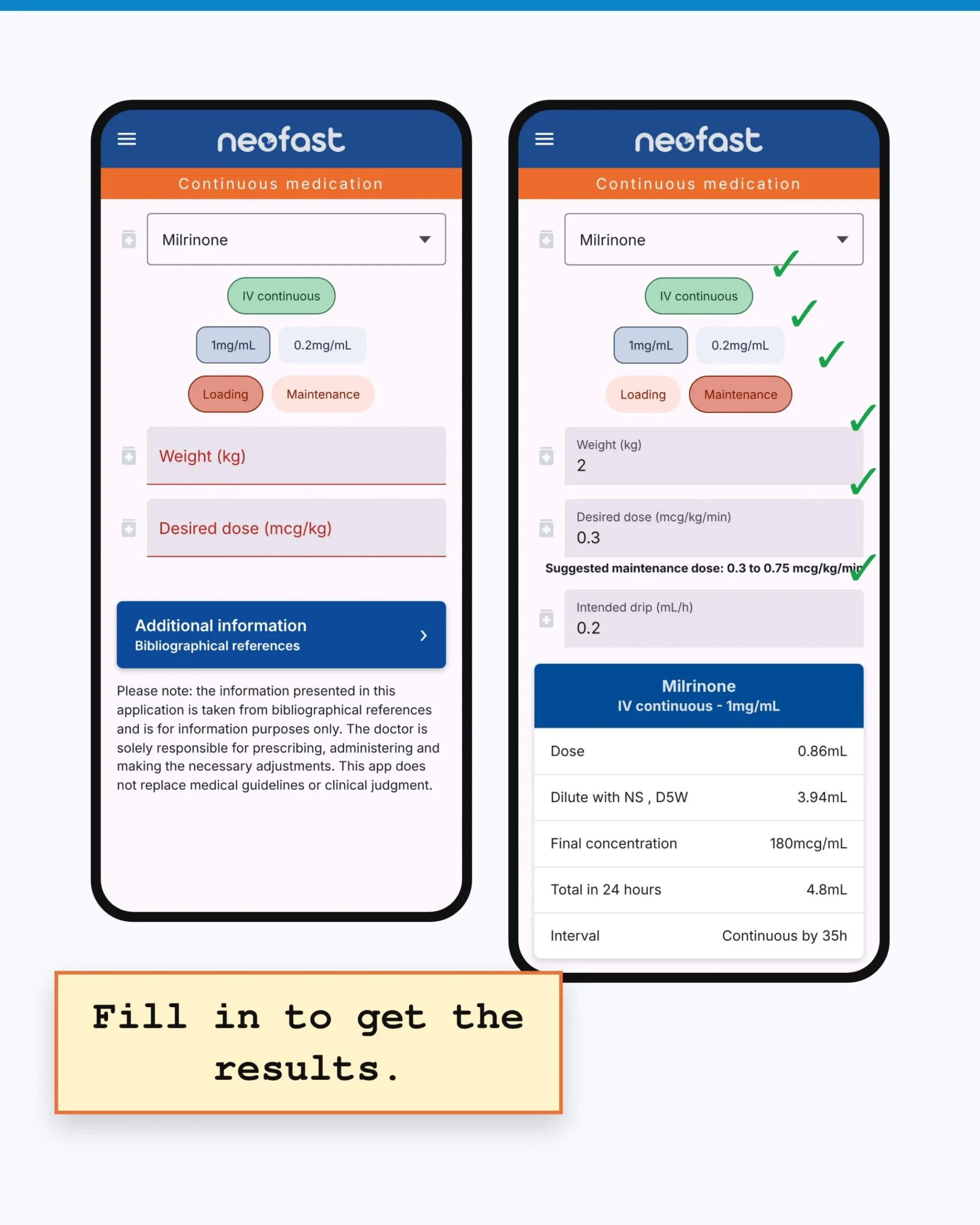
Continuous medication
Milrinone
- IV continuous
- 1mg/mL / 0.2mg/mL
- Loading / Maintenance
- Weight (kg)
- Desired dose (mcg/kg)
Additional information – Bibliographical references
Please note: the information presented in this application is taken from bibliographical references and is for information purposes only. The doctor is solely responsible for prescribing, administering and making the necessary adjustments. This app does not replace medical guidelines or clinical judgment.
Milrinone – filled example
- IV continuous
- 1mg/mL / 0.2mg/mL
- Loading / Maintenance
- Weight (kg): 2
- Desired dose (mcg/kg/min): 0.3
Suggested maintenance dose: 0.3 to 0.75 mcg/kg/min
- Intended drip (mL/h): 0.2
| Milrinone – IV continuous – 1mg/mL | |
|---|---|
| Dose | 0.86mL |
| Dilute with NS, D5W | 3.94mL |
| Final concentration | 180mcg/mL |
| Total in 24 hours | 4.8mL |
| Interval | Continuous by 35h |
Fill in to get the results.
Phases of Shock and Cellular Injury
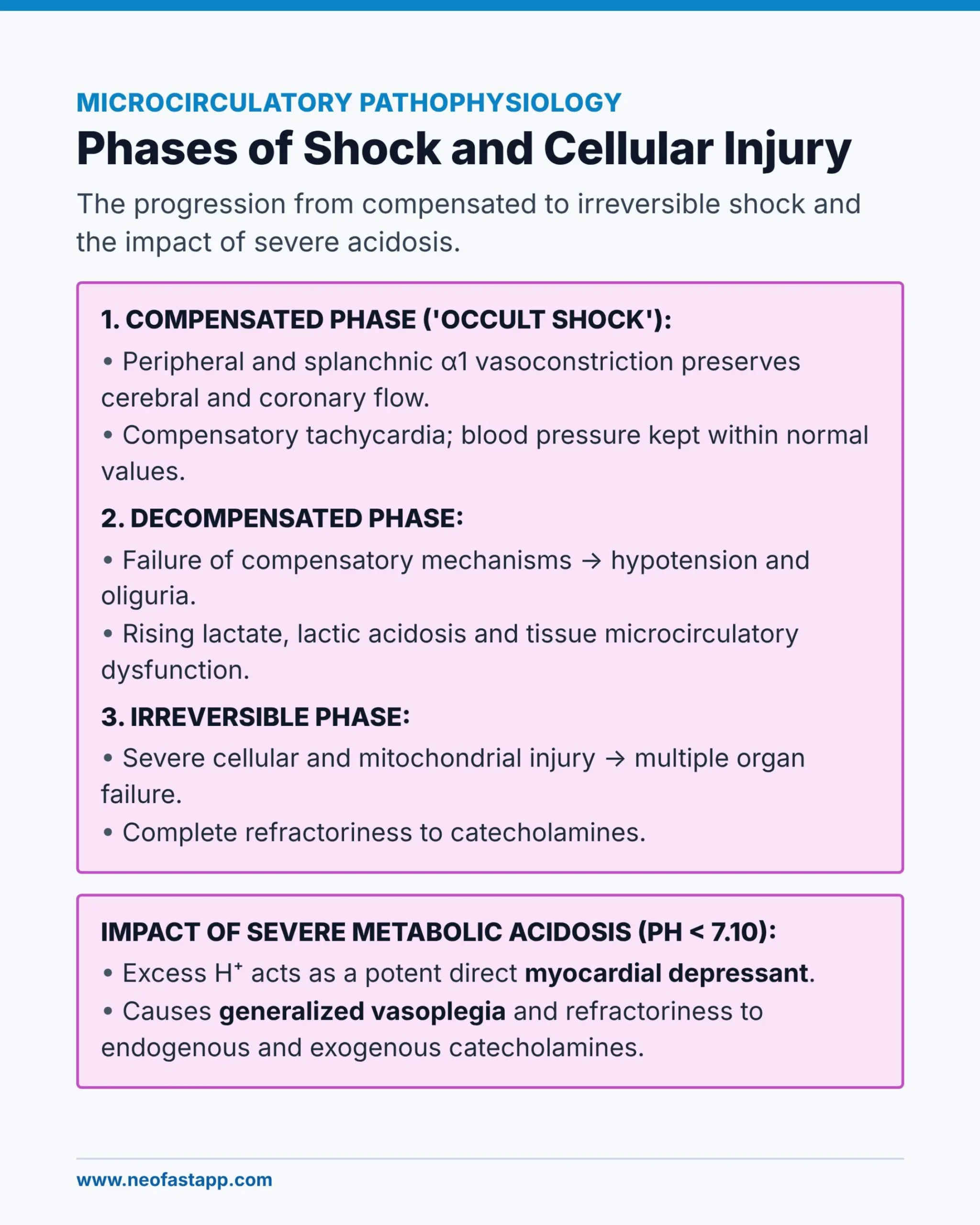
Microcirculatory Pathophysiology
The progression from compensated to irreversible shock and the impact of severe acidosis.
1. Compensated Phase (‘Occult Shock’)
- Peripheral and splanchnic α1 vasoconstriction preserves cerebral and coronary flow.
- Compensatory tachycardia; blood pressure kept within normal values.
2. Decompensated Phase
- Failure of compensatory mechanisms → hypotension and oliguria.
- Rising lactate, lactic acidosis and tissue microcirculatory dysfunction.
3. Irreversible Phase
- Severe cellular and mitochondrial injury → multiple organ failure.
- Complete refractoriness to catecholamines.
Impact of Severe Metabolic Acidosis (pH < 7.10)
- Excess H⁺ acts as a potent direct myocardial depressant.
- Causes generalized vasoplegia and refractoriness to endogenous and exogenous catecholamines.
Clinical Picture and Warning Signs
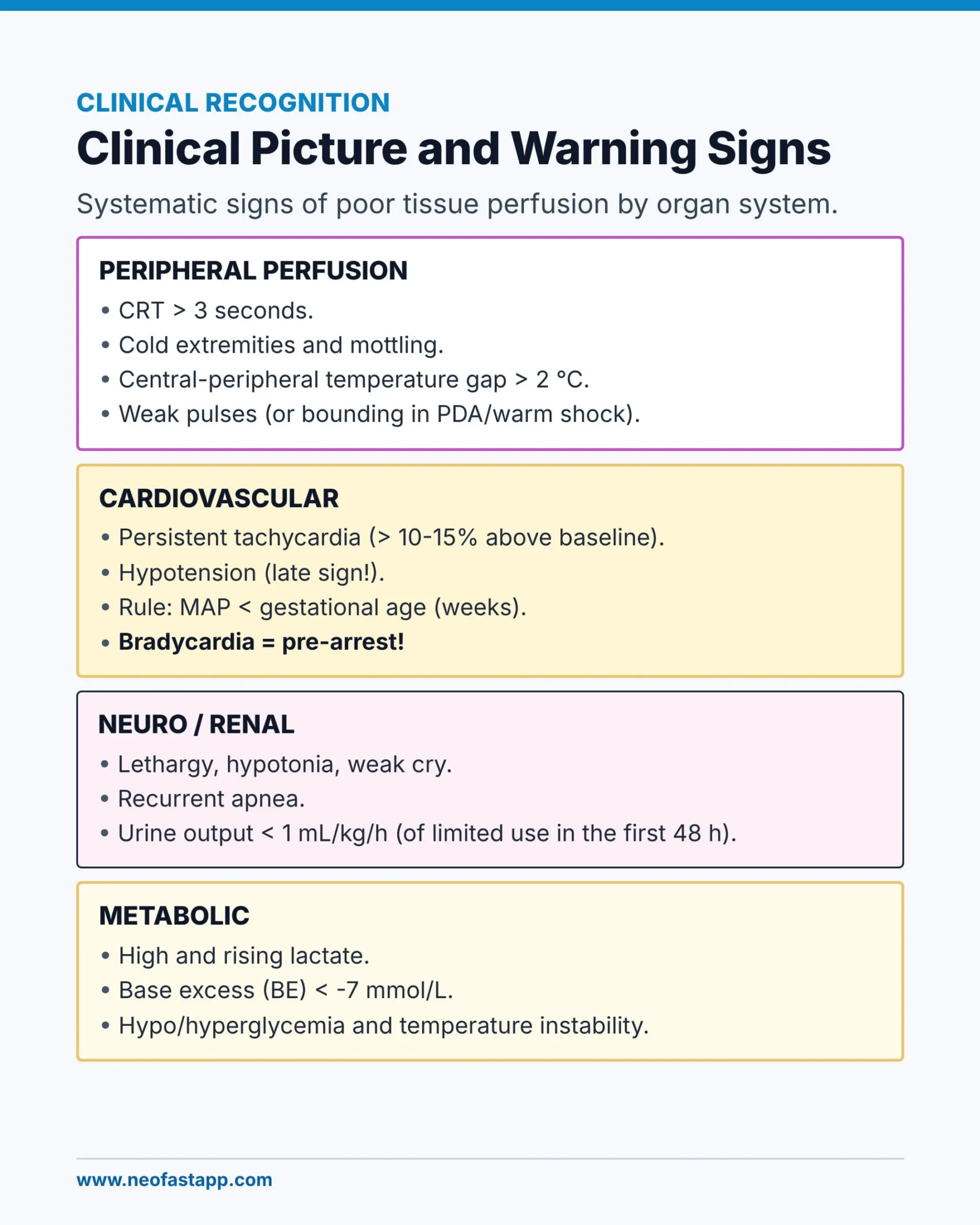
Clinical Recognition
Systematic signs of poor tissue perfusion by organ system.
Peripheral Perfusion
- CRT > 3 seconds.
- Cold extremities and mottling.
- Central-peripheral temperature gap > 2 °C.
- Weak pulses (or bounding in PDA/warm shock).
Cardiovascular
- Persistent tachycardia (> 10-15% above baseline).
- Hypotension (late sign!).
- Rule: MAP < gestational age (weeks).
- Bradycardia = pre-arrest!
Neuro / Renal
- Lethargy, hypotonia, weak cry.
- Recurrent apnea.
- Urine output < 1 mL/kg/h (of limited use in the first 48 h).
Metabolic
- High and rising lactate.
- Base excess (BE) < -7 mmol/L.
- Hypo/hyperglycemia and temperature instability.
Accuracy of Clinical Parameters and Tests
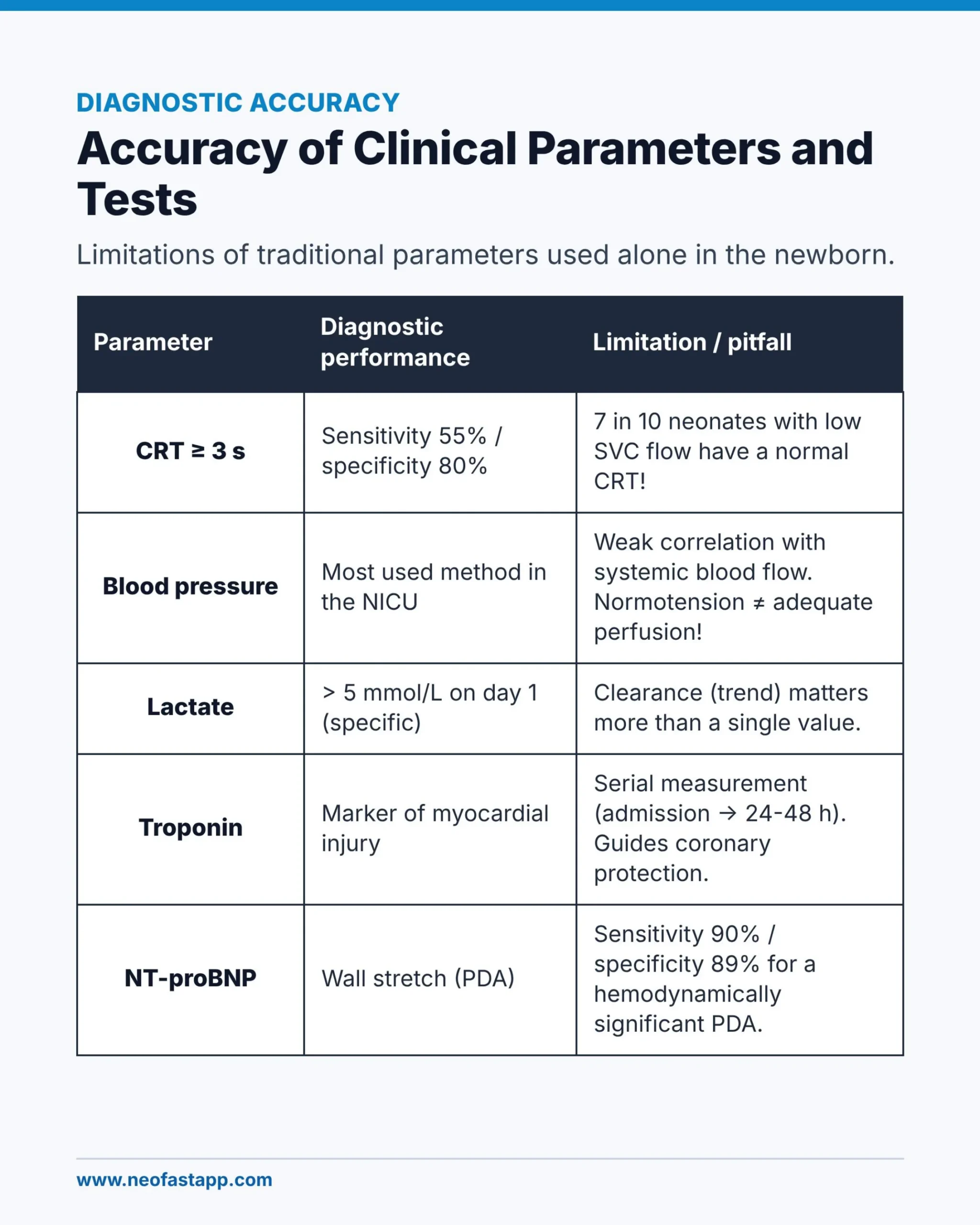
Diagnostic Accuracy
Limitations of traditional parameters used alone in the newborn.
| Parameter | Diagnostic performance | Limitation / pitfall |
|---|---|---|
| CRT ≥ 3 s | Sensitivity 55% / specificity 80% | 7 in 10 neonates with low SVC flow have a normal CRT! |
| Blood pressure | Most used method in the NICU | Weak correlation with systemic blood flow. Normotension ≠ adequate perfusion! |
| Lactate | > 5 mmol/L on day 1 (specific) | Clearance (trend) matters more than a single value. |
| Troponin | Marker of myocardial injury | Serial measurement (admission → 24-48 h). Guides coronary protection. |
| NT-proBNP | Wall stretch (PDA) | Sensitivity 90% / specificity 89% for a hemodynamically significant PDA. |
Monitoring Tools in the NICU

Advanced Monitoring
Invasive and non-invasive methods of hemodynamic assessment compared.
| Method | What it tells you | Advantage / limitation |
|---|---|---|
| Invasive BP | Continuous BP; access for blood gases | Gold standard. Risk of thrombosis and damped waveform. |
| NIRS (rSO₂) | Tissue oxygenation (brain, kidney, splanchnic) | Continuous (ideal range: 55-85%). Mean and trend. |
| ScvO₂ | Global O₂ extraction (DO₂/VO₂ balance) | Target ≥ 70%. Umbilical catheter position changes the value. |
| TnECHO / POCUS | Preload, contractility, SVR, shunts, PH | Defines the hemodynamic phenotype. Requires training. |
POCUS in Shock: the SAFE-R Protocol
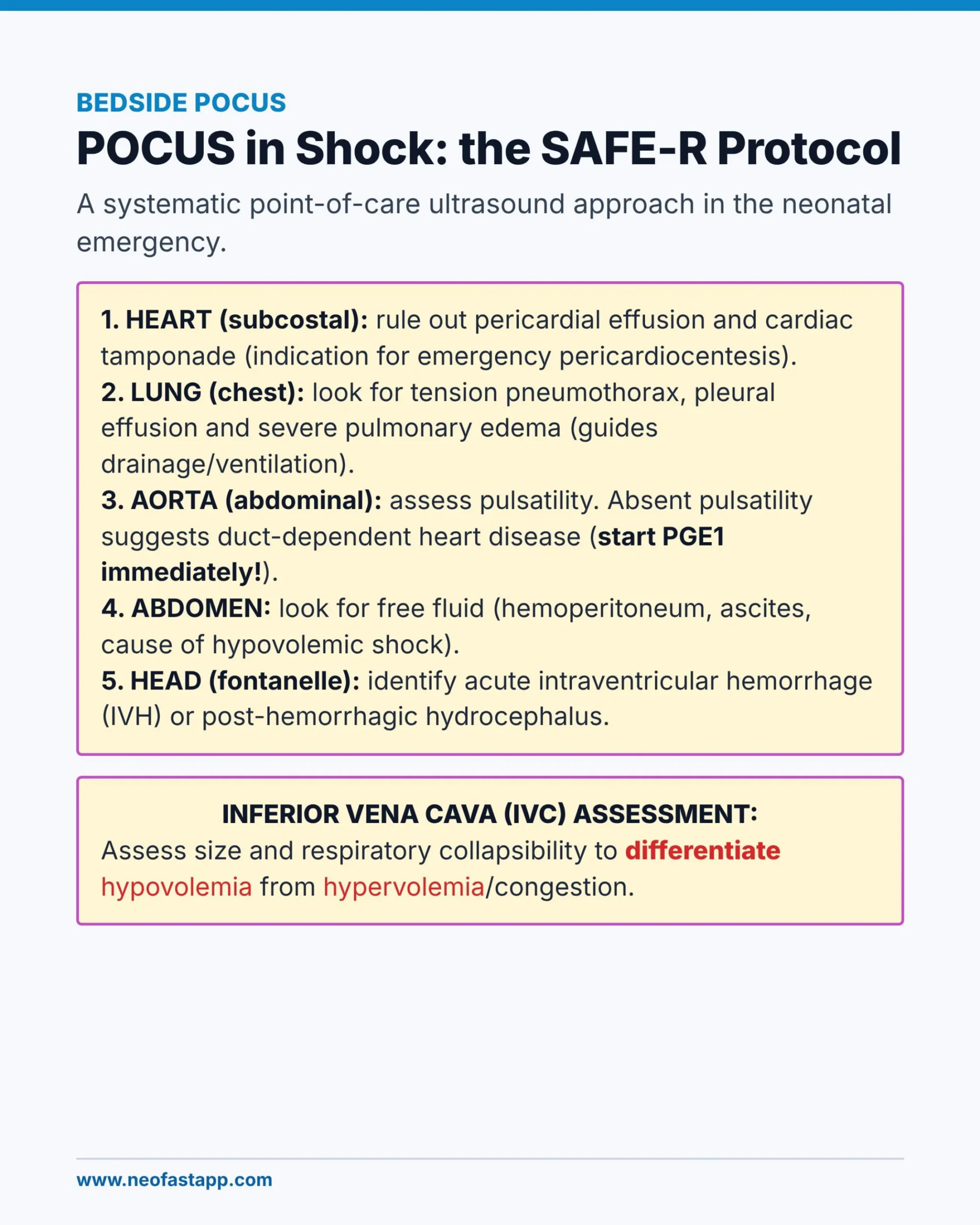
Bedside POCUS
A systematic point-of-care ultrasound approach in the neonatal emergency.
- Heart (subcostal): rule out pericardial effusion and cardiac tamponade (indication for emergency pericardiocentesis).
- Lung (chest): look for tension pneumothorax, pleural effusion and severe pulmonary edema (guides drainage/ventilation).
- Aorta (abdominal): assess pulsatility. Absent pulsatility suggests duct-dependent heart disease (start PGE1 immediately!).
- Abdomen: look for free fluid (hemoperitoneum, ascites, cause of hypovolemic shock).
- Head (fontanelle): identify acute intraventricular hemorrhage (IVH) or post-hemorrhagic hydrocephalus.
Inferior Vena Cava (IVC) Assessment
Assess size and respiratory collapsibility to differentiate hypovolemia from hypervolemia/congestion.
TnECHO Parameters in Neonatal Shock
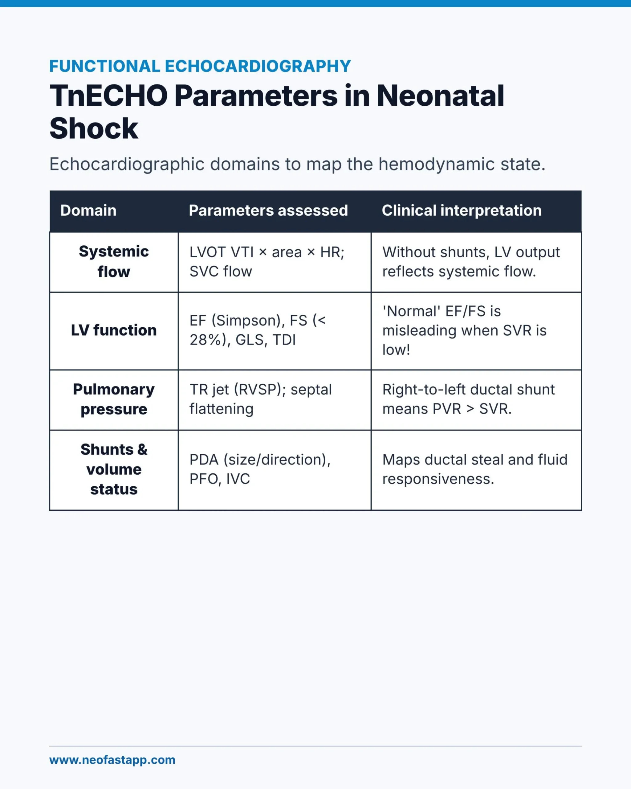
Functional Echocardiography
Echocardiographic domains to map the hemodynamic state.
| Domain | Parameters assessed | Clinical interpretation |
|---|---|---|
| Systemic flow | LVOT VTI × area × HR; SVC flow | Without shunts, LV output reflects systemic flow. |
| LV function | EF (Simpson), FS (< 28%), GLS, TDI | ‘Normal’ EF/FS is misleading when SVR is low! |
| Pulmonary pressure | TR jet (RVSP); septal flattening | Right-to-left ductal shunt means PVR > SVR. |
| Shunts & volume status | PDA (size/direction), PFO, IVC | Maps ductal steal and fluid responsiveness. |
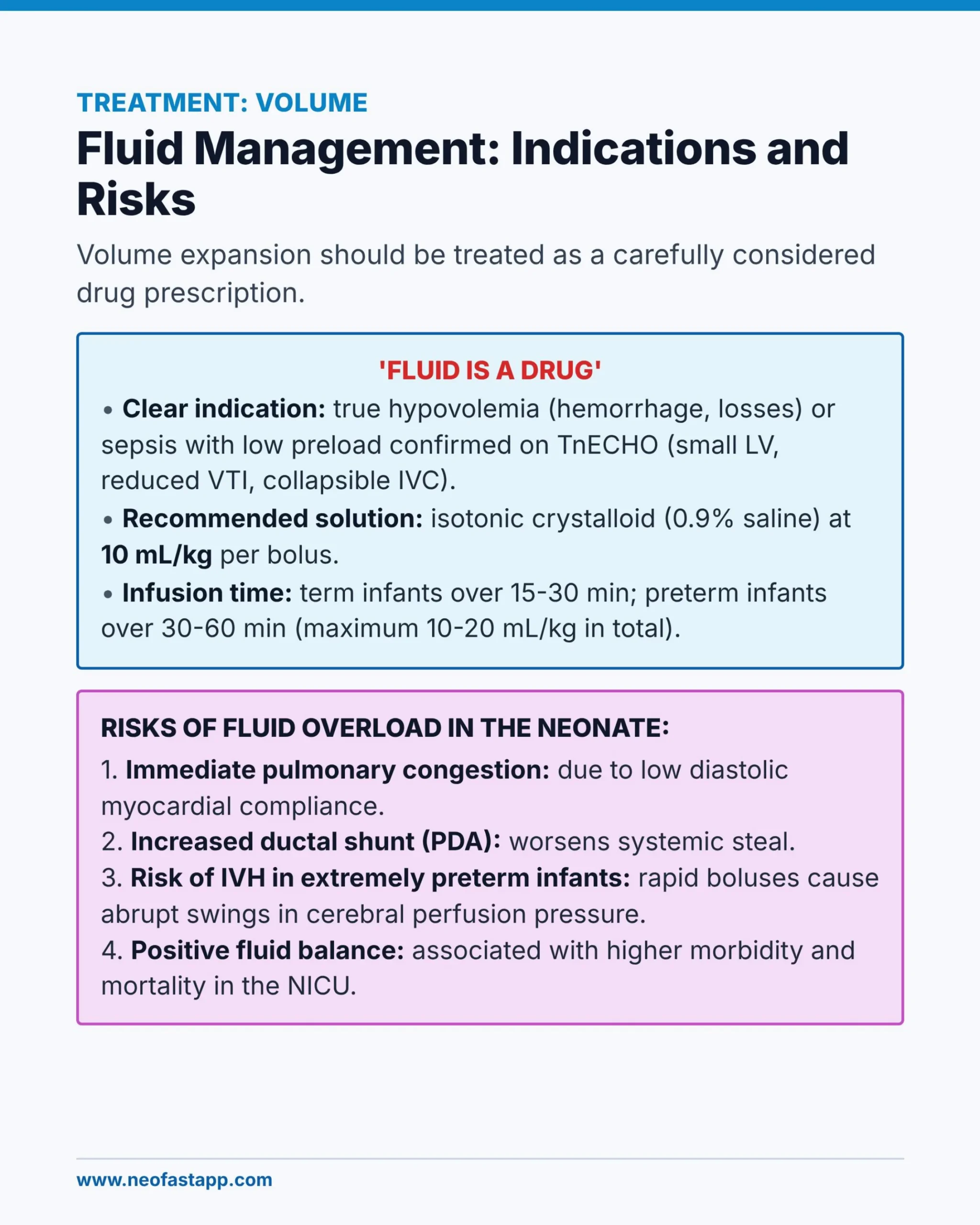
Fluid Management: Indications and Risks
Treatment: Volume
Volume expansion should be treated as a carefully considered drug prescription.
‘Fluid is a Drug’
- Clear indication: true hypovolemia (hemorrhage, losses) or sepsis with low preload confirmed on TnECHO (small LV, reduced VTI, collapsible IVC).
- Recommended solution: isotonic crystalloid (0.9% saline) at 10 mL/kg per bolus.
- Infusion time: term infants over 15-30 min; preterm infants over 30-60 min (maximum 10-20 mL/kg in total).
Risks of Fluid Overload in the Neonate
- Immediate pulmonary congestion: due to low diastolic myocardial compliance.
- Increased ductal shunt (PDA): worsens systemic steal.
- Risk of IVH in extremely preterm infants: rapid boluses cause abrupt swings in cerebral perfusion pressure.
- Positive fluid balance: associated with higher morbidity and mortality in the NICU.
Vasoactive Drugs and Corticosteroids

Pharmacotherapy
Choice of vasoactive drug guided by the newborn’s hemodynamic phenotype.
| Drug & dose | Main mechanism | Predominant effect & cautions |
|---|---|---|
| Dobutamine 5-20 mcg/kg/min | Inotrope / inodilator (β1 > β2) | ++++ inotropy, ++ vasodilation. Caution: tachycardia and ↑ O₂ consumption. |
| Epinephrine 0.05-1 mcg/kg/min | Inotrope / vasopressor (β1/β2 at low doses; α1 at high doses) | Potent inotrope. High doses cause vasoconstriction. Raises lactate/glucose. |
| Norepinephrine 0.05-1 mcg/kg/min | Potent vasopressor (α1 >> β1) | ++++ vasoconstriction. Requires corrected volume status. Ideal in warm shock. |
| Milrinone 0.25-0.75 mcg/kg/min | Inodilator (PDE3 inhibitor) | ++++ inotropy/lusitropy. Lowers PVR/SVR. Watch for hypotension. |
Corticosteroids
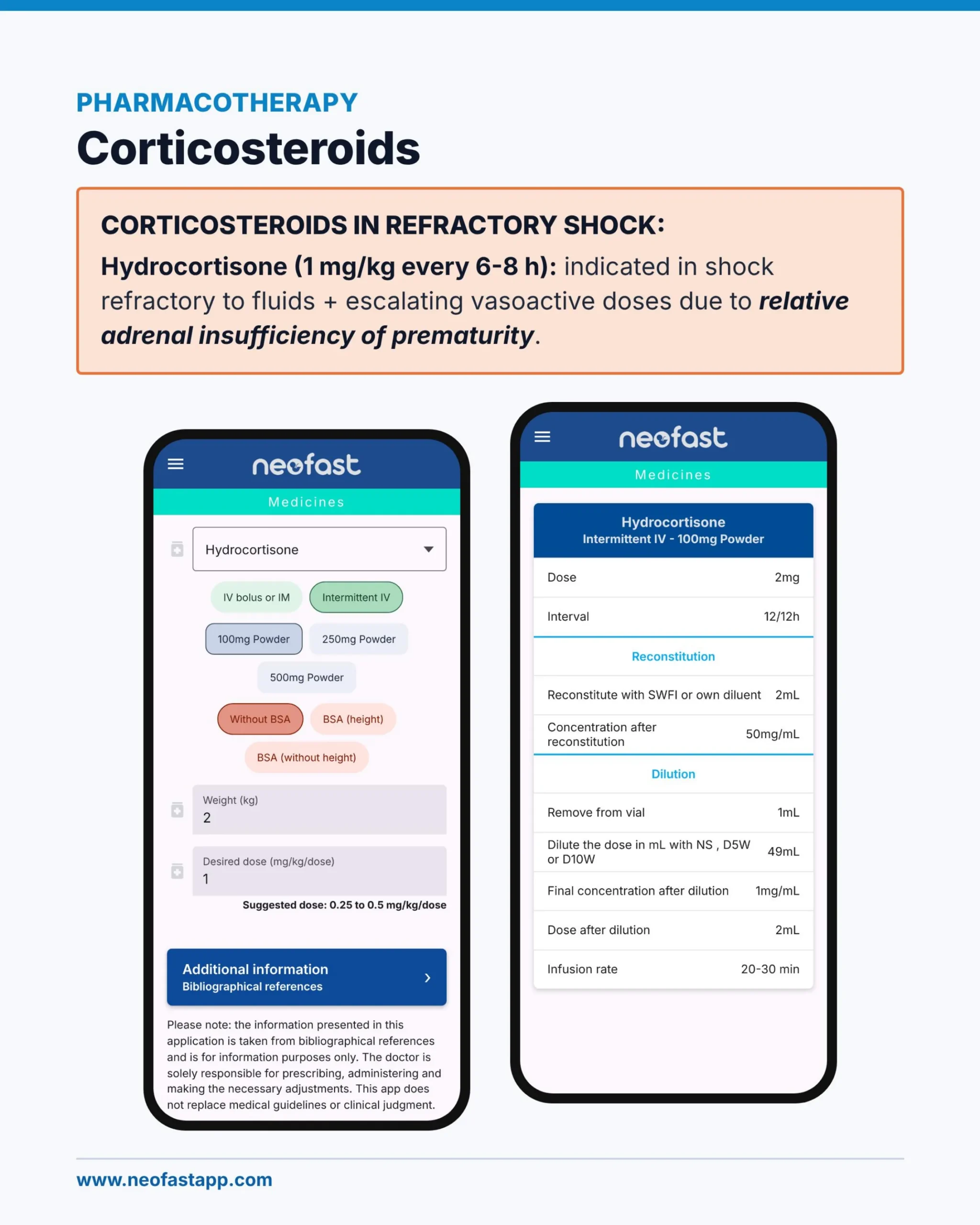
Pharmacotherapy: Corticosteroids
CORTICOSTEROIDS IN REFRACTORY SHOCK:
Hydrocortisone (1 mg/kg every 6-8 h): indicated in shock refractory to fluids + escalating vasoactive doses due to relative adrenal insufficiency of prematurity.
Neofast App – Medicines Screen
- Drug: Hydrocortisone
- Route options: IV bolus or IM, Intermittent IV (selected)
- Formulation options: 100mg Powder (selected), 250mg Powder, 500mg Powder
- BSA options: Without BSA (selected), BSA (height), BSA (without height)
- Weight (kg): 2
- Desired dose (mg/kg/dose): 1
- Suggested dose: 0.25 to 0.5 mg/kg/dose
- Additional information / Bibliographical references (link)
Please note: the information presented in this application is taken from bibliographical references and is for information purposes only. The doctor is solely responsible for prescribing, administering and making the necessary adjustments. This app does not replace medical guidelines or clinical judgment.
Neofast App – Hydrocortisone Detail (Intermittent IV – 100mg Powder)
| Parameter | Value |
|---|---|
| Dose | 2mg |
| Interval | 12/12h |
Reconstitution
| Reconstitute with SWFI or own diluent | 2mL |
| Concentration after reconstitution | 50mg/mL |
Dilution
| Remove from vial | 1mL |
| Dilute the dose in mL with NS, D5W or D10W | 49mL |
| Final concentration after dilution | 1mg/mL |
| Dose after dilution | 2mL |
| Infusion rate | 20-30 min |
Final Summary – Key Points

Final Summary
Key Points ⚠️
- Neonatal myocardium: depends on HR, needs Ca²⁺ and tolerates afterload poorly.
- Normal BP ≠ adequate perfusion: monitor microcirculation signs and tissue O₂ delivery.
- Lactate and BE: value the trend over time (clearance), not a single value.
- Hemodynamic phenotyping: management should be guided by TnECHO / POCUS.
- Fluid is a drug: 10 mL/kg boluses with careful reassessment (caution in preterm infants).
- Choice of vasoactive drug: pick it according to the pathophysiology (SVR vs. contractility).
- Refractory shock: hydrocortisone, TSH/free T4, and re-investigate PDA, PPHN and heart disease.
- Beware of acidosis: do not use sodium bicarbonate routinely, and do not deplete K⁺ during the acidotic phase.
Neofast
Faster shifts — Download on the App Store
Easier shifts — Get it on Google Play
Check it out!
Frequently asked questions
What is the difference between warm shock and cold shock in neonatal sepsis?
Warm shock (↓SVR) presents with warm extremities, rapid ‘flash’ CRT, bounding pulses and low diastolic BP, with a hyperdynamic, underfilled heart on TnECHO; it is treated with a vasopressor alone (norepinephrine or vasopressin). Cold shock (↑↑SVR) presents with cold extremities, CRT >3 s, weak pulses and late hypotension, with LV or biventricular systolic dysfunction and reactive pulmonary hypertension; it is treated with an inotrope/inodilator (epinephrine or dobutamine/milrinone).
What are the recommended doses of vasoactive drugs in neonatal shock?
Dobutamine 5-20 mcg/kg/min (inotrope/inodilator, caution with tachycardia); epinephrine 0.05-1 mcg/kg/min (potent inotrope, raises lactate/glucose at high doses); norepinephrine 0.05-1 mcg/kg/min (potent vasopressor, ideal in warm shock, requires corrected volume status); milrinone 0.25-0.75 mcg/kg/min (inodilator/PDE3 inhibitor, lowers PVR/SVR, watch for hypotension).
How should fluid boluses be given in neonatal shock and what are the risks?
Use isotonic crystalloid (0.9% saline) at 10 mL/kg per bolus, infused over 15-30 min in term infants and 30-60 min in preterm infants, with a maximum total of 10-20 mL/kg, only when true hypovolemia or low preload is confirmed on TnECHO. Risks of overload include immediate pulmonary congestion, increased PDA shunt/systemic steal, IVH in extremely preterm infants from rapid boluses, and higher morbidity/mortality with positive fluid balance.
When is hydrocortisone indicated in neonatal shock and at what dose?
Hydrocortisone 1 mg/kg every 6-8 h is indicated for shock refractory to fluids plus escalating vasoactive doses, addressing relative adrenal insufficiency of prematurity.
Why is blood pressure an unreliable marker of perfusion in neonatal shock?
Blood pressure is the most used NICU monitoring method but has weak correlation with systemic blood flow, so normotension does not equal adequate perfusion; hypotension is also a late sign, and the rule of thumb is that MAP below gestational age in weeks signals concern, while bradycardia indicates pre-arrest.