


Key takeaways
- Hypoxic-ischemic encephalopathy is the most common cause of neonatal seizures in full-term newborns, typically presenting within the first 6 to 12 hours of life.
- Most neonatal seizures are purely electrographic with no visible motor manifestations, and after anticonvulsant treatment, motor symptoms may disappear while equally harmful electrical seizures continue (electroclinical decoupling).
- First-line treatment is phenobarbital 20 mg/kg IV over 10–20 minutes, repeatable at 10–20 mg/kg up to a maximum of 40 mg/kg total, with maintenance of 3–5 mg/kg/day.
- Levetiracetam (second-line, 40–60 mg/kg IV over 15 min) has an excellent safety profile with no cardiorespiratory depression, while phenytoin (second-line, 15–20 mg/kg IV, <1 mg/kg/min) requires cardiac monitoring and a large-bore IV line due to necrosis risk.
- cEEG should be maintained for 24 hours after the last seizure before considering complete control, and in acute induced seizures (asphyxia, perinatal ischemic stroke, isolated lobar hemorrhage, controlled infection) with resolved electrical seizures, anticonvulsants should be discontinued before discharge since there is no benefit to long-term maintenance.
References
- Meijler G, Mohammad K (Eds.). Neonatal Brain Injury: An Illustrated Guide for Clinicians Counselling Parents and Caregivers. Springer, 2024. Open Access.
- Glass HC et al. Safety of early discontinuation of anticonvulsants after symptomatic acute neonatal seizures. JAMA Neurology, 2021.
- Glass HC et al., JAMA Neurology, 2021.
- Meijler & Mohammad (Eds.), Neonatal Brain Injury, Springer 2024 (Open Access)
Neonatal seizures
Neonatal seizures
Recognize, monitor, and treat within the first few hours.
Pathophysiology: Why does a newborn’s brain convulse so easily?
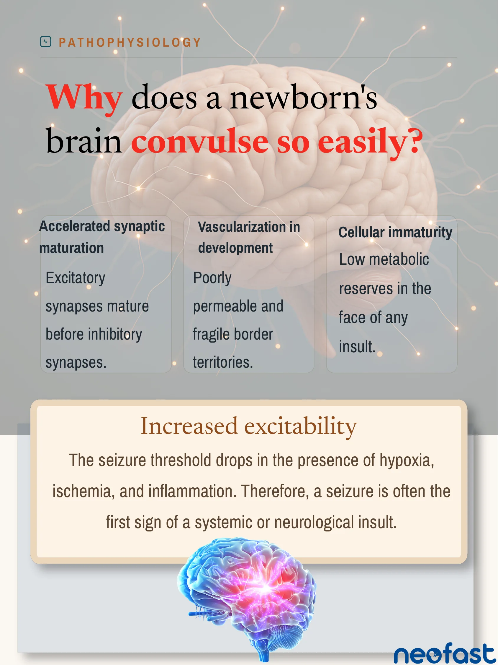
Why does a newborn’s brain convulse so easily?
- Accelerated synaptic maturation: Excitatory synapses mature before inhibitory synapses.
- Vascularization in development: Poorly permeable and fragile border territories.
- Cellular immaturity: Low metabolic reserves in the face of any insult.
Increased excitability
The seizure threshold drops in the presence of hypoxia, ischemia, and inflammation. Therefore, a seizure is often the first sign of a systemic or neurological insult.
Etiology · Term Newborn: The three main causes
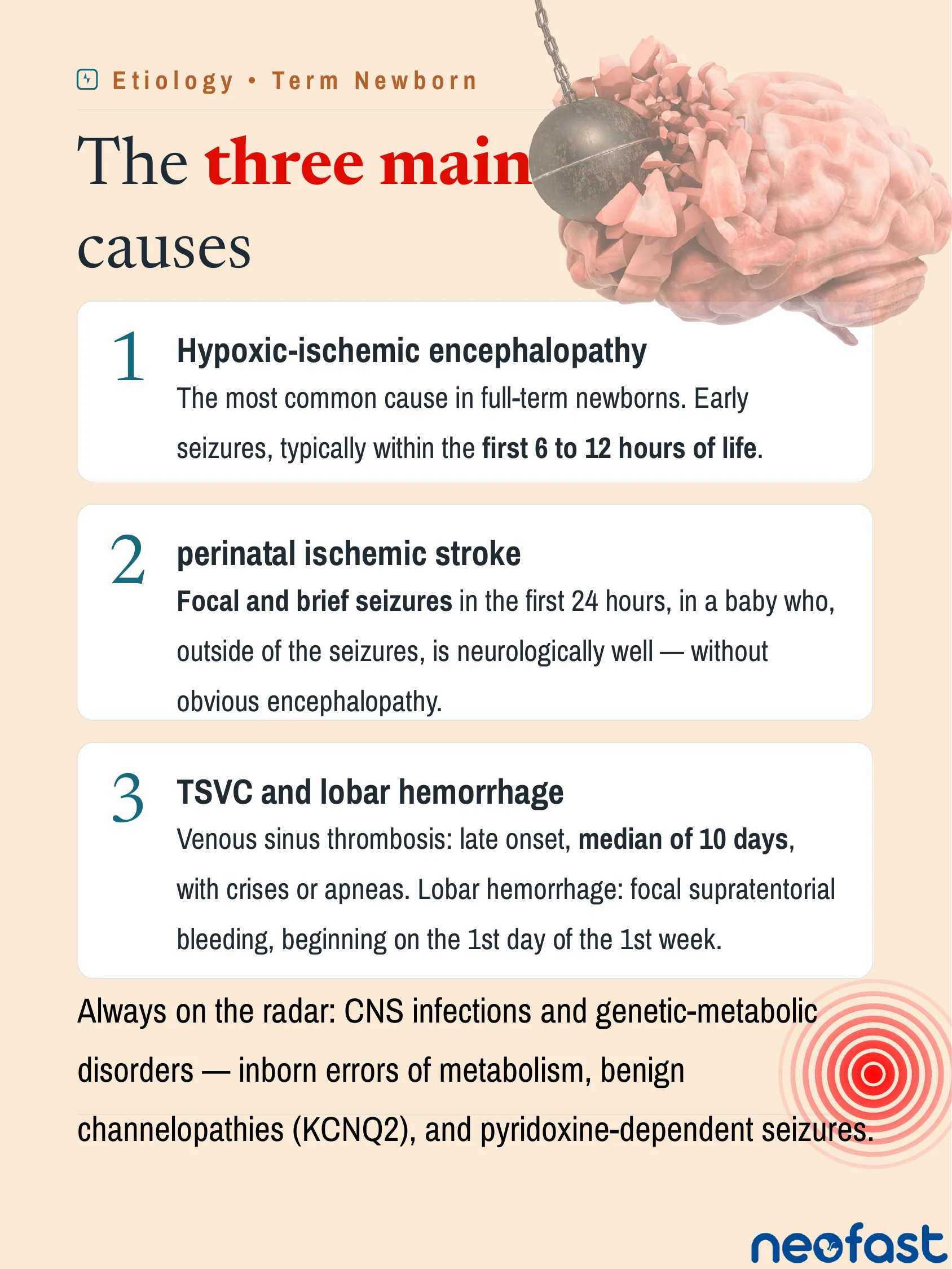
The three main causes
- Hypoxic-ischemic encephalopathy
The most common cause in full-term newborns. Early seizures, typically within the first 6 to 12 hours of life.
- Perinatal ischemic stroke
Focal and brief seizures in the first 24 hours, in a baby who, outside of the seizures, is neurologically well — without obvious encephalopathy.
- TSVC and lobar hemorrhage
Venous sinus thrombosis: late onset, median of 10 days, with crises or apneas. Lobar hemorrhage: focal supratentorial bleeding, beginning on the 1st day of the 1st week.
Always on the radar: CNS infections and genetic-metabolic disorders — inborn errors of metabolism, benign channelopathies (KCNQ2), and pyridoxine-dependent seizures.
Differential Diagnosis: The age at birth guides the diagnosis

The age at birth guides the diagnosis.
| Timing | Diagnosis | Description |
|---|---|---|
| 6–12 h | Hypoxic-ischemic encephalopathy | Most common cause in full-term newborns |
| < 24 h | Perinatal ischemic stroke | Focal and brief seizures, baby doing well between them. |
| Day 1 | Lobar hemorrhage | Mechanical delivery or coagulation disorder |
| Days 4–6 | Rotavirus | The classic “fifth day fits” |
| ~Day 10 | Venous sinus thrombosis | Seizures or apneas — median of 10 days |
| ~Day 16 | Enterovirus | Late-window viral encephalitis |
| 16th–19th day | Herpes simples (HSV) | Lethargy + seizures. Don’t wait for the gallbladders. |
| Late | Bacterial meningitis | GBS or E. coli |
The fallacy of the clinical examination: Most crises are silent
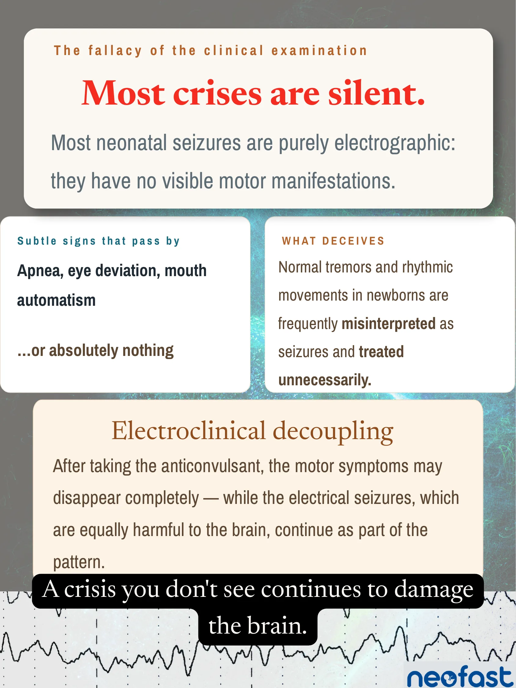
Most crises are silent.
Most neonatal seizures are purely electrographic: they have no visible motor manifestations.
Subtle signs that pass by
Apnea, eye deviation, mouth automatism …or absolutely nothing
What deceives
Normal tremors and rhythmic movements in newborns are frequently misinterpreted as seizures and treated unnecessarily.
Electroclinical decoupling
After taking the anticonvulsant, the motor symptoms may disappear completely — while the electrical seizures, which are equally harmful to the brain, continue as part of the pattern.
A crisis you don’t see continues to damage the brain.
Monitoring: cEEG or aEEG? Both — for different things
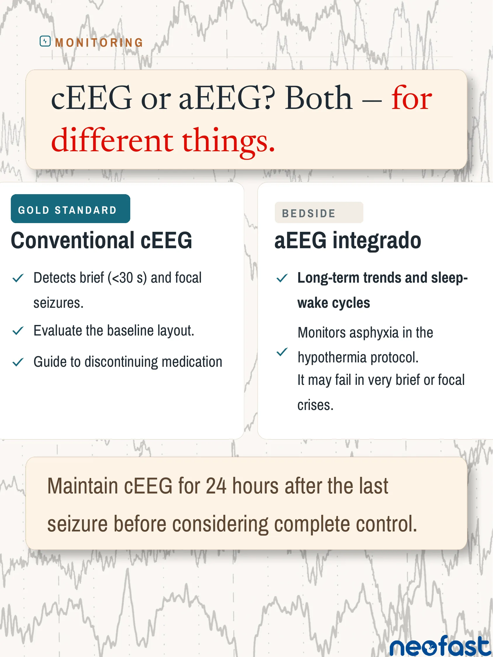
cEEG or aEEG? Both — for different things.
Conventional cEEG (Gold standard)
- Detects brief (<30 s) and focal seizures.
- Evaluate the baseline layout.
- Guide to discontinuing medication
aEEG integrado (Bedside)
- Long-term trends and sleep-wake cycles
- Monitors asphyxia in the hypothermia protocol.
It may fail in very brief or focal crises.
Maintain cEEG for 24 hours after the last seizure before considering complete control.
Management · Step 1: Stabilize before discontinuing anticonvulsants
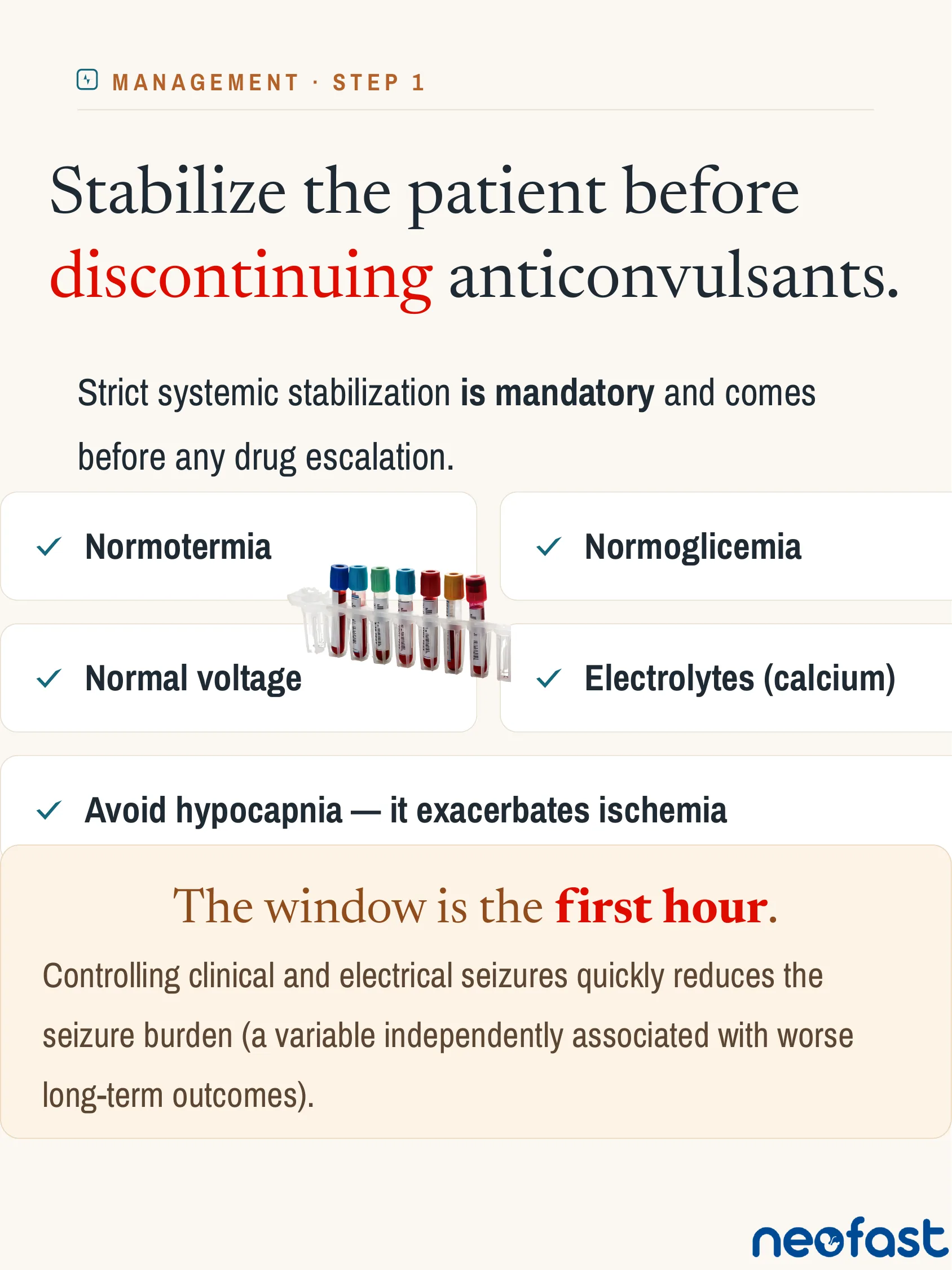
Stabilize the patient before discontinuing anticonvulsants.
Strict systemic stabilization is mandatory and comes before any drug escalation.
- Normotermia
- Normoglicemia
- Normal voltage
- Electrolytes (calcium)
- Avoid hypocapnia — it exacerbates ischemia
The window is the first hour.
Controlling clinical and electrical seizures quickly reduces the seizure burden (a variable independently associated with worse long-term outcomes).
Management · Step 2: The staircase: first and second row
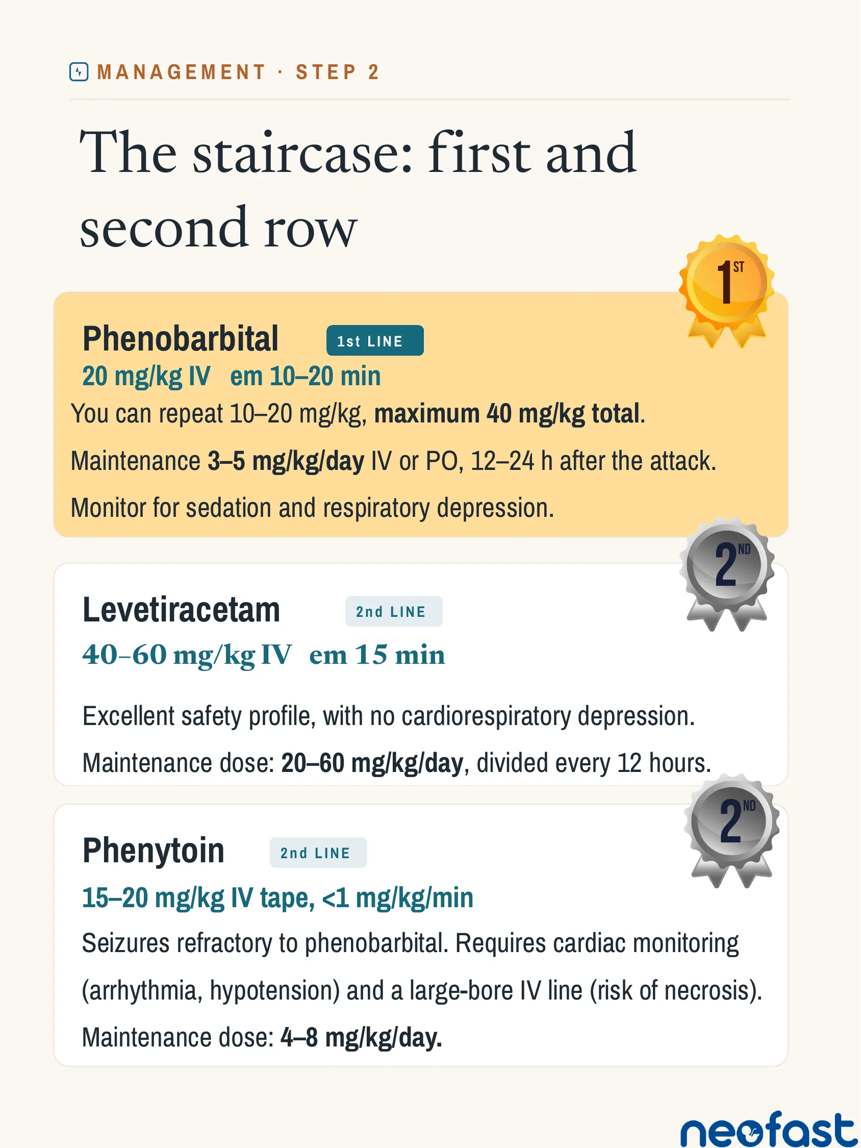
The staircase: first and second row
Phenobarbital (1st LINE)
20 mg/kg IV em 10–20 min
You can repeat 10–20 mg/kg, maximum 40 mg/kg total.
Maintenance 3–5 mg/kg/day IV or PO, 12–24 h after the attack.
Monitor for sedation and respiratory depression.
Levetiracetam (2nd LINE)
40–60 mg/kg IV em 15 min
Excellent safety profile, with no cardiorespiratory depression.
Maintenance dose: 20–60 mg/kg/day, divided every 12 hours.
Phenytoin (2nd LINE)
15–20 mg/kg IV tape, <1 mg/kg/min
Seizures refractory to phenobarbital. Requires cardiac monitoring (arrhythmia, hypotension) and a large-bore IV line (risk of necrosis).
Maintenance dose: 4–8 mg/kg/day.
Management · Step 3: Refractory and specific cases
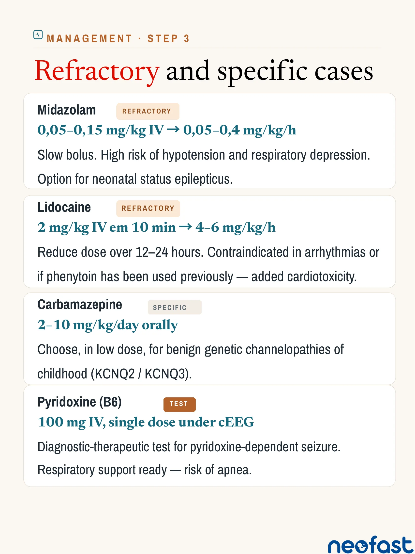
Management · Step 3
Refractory and specific cases
- Midazolam (Refractory)
0,05–0,15 mg/kg IV → 0,05–0,4 mg/kg/h
Slow bolus. High risk of hypotension and respiratory depression.
Option for neonatal status epilepticus.
- Lidocaine (Refractory)
2 mg/kg IV em 10 min → 4–6 mg/kg/h
Reduce dose over 12–24 hours. Contraindicated in arrhythmias or if phenytoin has been used previously — added cardiotoxicity.
- Carbamazepine (Specific)
2–10 mg/kg/day orally
Choose, in low dose, for benign genetic channelopathies of childhood (KCNQ2 / KCNQ3).
- Pyridoxine (B6) (Test)
100 mg IV, single dose under cEEG
Diagnostic-therapeutic test for pyridoxine-dependent seizure. Respiratory support ready — risk of apnea.
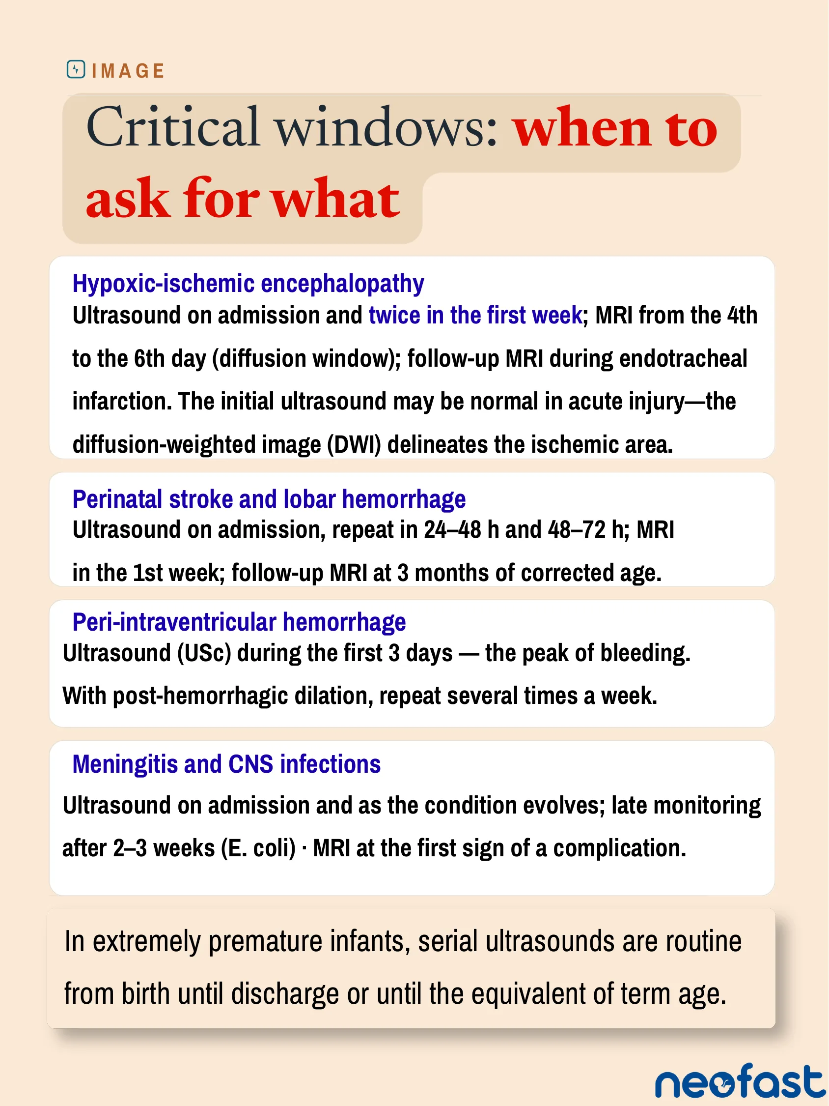
Critical windows: when to ask for what
Image
Critical windows: when to ask for what
- Hypoxic-ischemic encephalopathy
Ultrasound on admission and twice in the first week; MRI from the 4th to the 6th day (diffusion window); follow-up MRI during endotracheal infarction. The initial ultrasound may be normal in acute injury—the diffusion-weighted image (DWI) delineates the ischemic area.
- Perinatal stroke and lobar hemorrhage
Ultrasound on admission, repeat in 24–48 h and 48–72 h; MRI in the 1st week; follow-up MRI at 3 months of corrected age.
- Peri-intraventricular hemorrhage
Ultrasound (USc) during the first 3 days — the peak of bleeding. With post-hemorrhagic dilation, repeat several times a week.
- Meningitis and CNS infections
Ultrasound on admission and as the condition evolves; late monitoring after 2–3 weeks (E. coli) · MRI at the first sign of a complication.
In extremely premature infants, serial ultrasounds are routine from birth until discharge or until the equivalent of term age.
In practice: NeoFast dosing calculator
In Practice
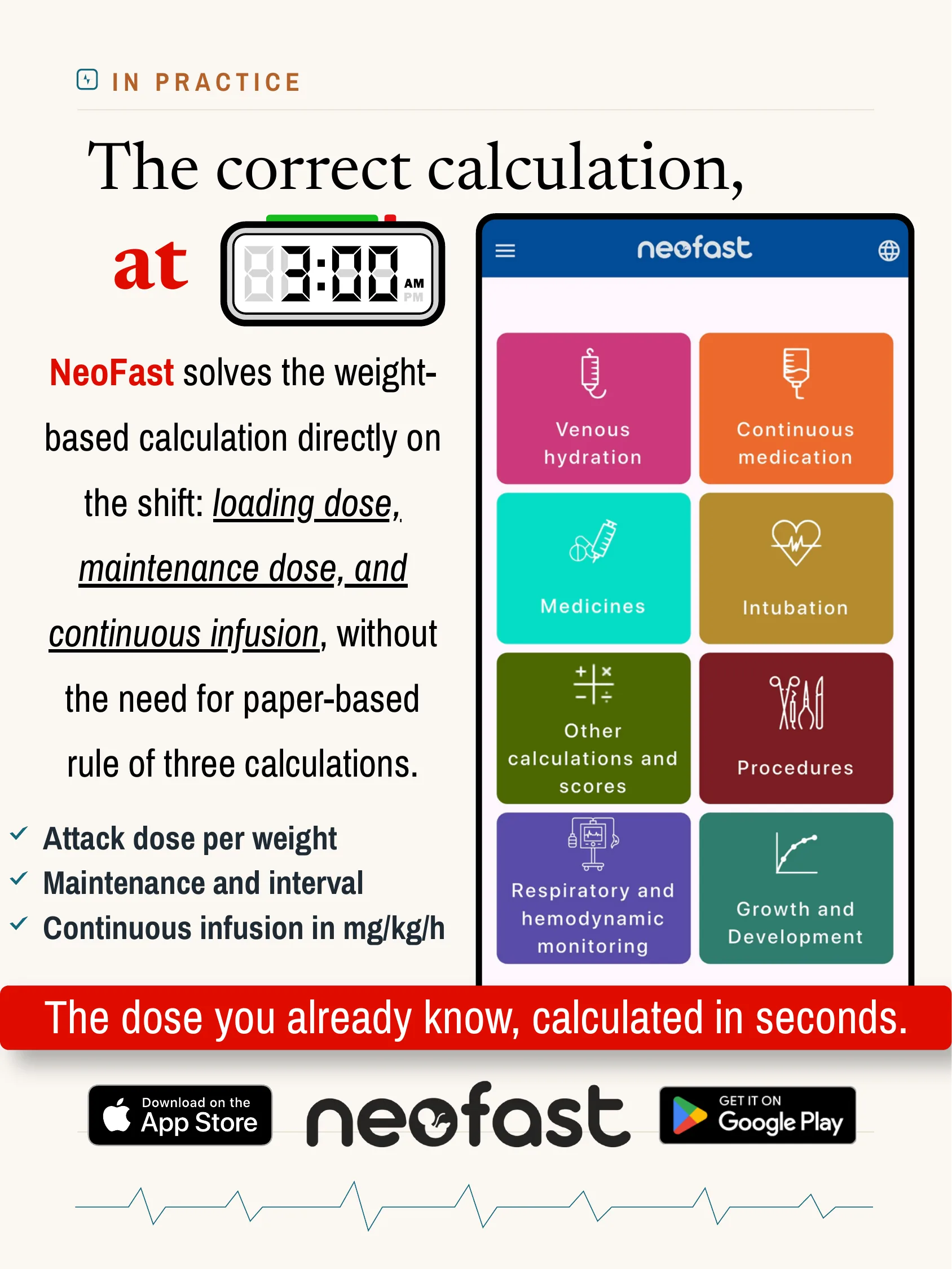
The correct calculation, at 3:00 AM
NeoFast solves the weight-based calculation directly on the shift: loading dose, maintenance dose, and continuous infusion, without the need for paper-based rule of three calculations.
- Attack dose per weight
- Maintenance and interval
- Continuous infusion in mg/kg/h
The dose you already know, calculated in seconds.
- Venous hydration
- Continuous medication
- Medicines
- Intubation
- Other calculations and scores
- Procedures
- Respiratory and hemodynamic monitoring
- Growth and Development
For the duty: Two things that almost everyone gets wrong
For The Duty
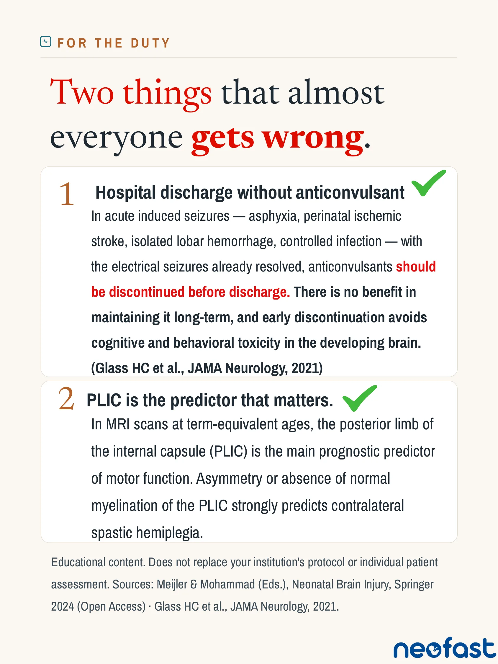
Two things that almost everyone gets wrong.
- Hospital discharge without anticonvulsant ✔
In acute induced seizures — asphyxia, perinatal ischemic stroke, isolated lobar hemorrhage, controlled infection — with the electrical seizures already resolved, anticonvulsants should be discontinued before discharge. There is no benefit in maintaining it long-term, and early discontinuation avoids cognitive and behavioral toxicity in the developing brain. (Glass HC et al., JAMA Neurology, 2021)
- PLIC is the predictor that matters. ✔
In MRI scans at term-equivalent ages, the posterior limb of the internal capsule (PLIC) is the main prognostic predictor of motor function. Asymmetry or absence of normal myelination of the PLIC strongly predicts contralateral spastic hemiplegia.
Educational content. Does not replace your institution’s protocol or individual patient assessment. Sources: Meijler & Mohammad (Eds.), Neonatal Brain Injury, Springer 2024 (Open Access) · Glass HC et al., JAMA Neurology, 2021.
Frequently asked questions
What is the most common cause of neonatal seizures in full-term newborns and when do they occur?
Hypoxic-ischemic encephalopathy is the most common cause in full-term newborns, with early seizures typically occurring within the first 6 to 12 hours of life.
What is the first-line drug for neonatal seizures and what is the dosing?
Phenobarbital is first-line: 20 mg/kg IV over 10–20 minutes, which can be repeated at 10–20 mg/kg up to a maximum of 40 mg/kg total, with a maintenance dose of 3–5 mg/kg/day IV or PO given 12–24 hours after the attack, while monitoring for sedation and respiratory depression.
Why can neonatal seizures be missed on clinical examination alone?
Most neonatal seizures are purely electrographic with no visible motor manifestations; subtle signs like apnea, eye deviation, or mouth automatisms may occur, or there may be no visible signs at all, and normal newborn tremors are often misinterpreted as seizures and treated unnecessarily.
Should cEEG or aEEG be used to monitor neonatal seizures?
Both are used for different purposes: conventional cEEG (gold standard) detects brief (<30 s) and focal seizures, evaluates baseline pattern, and guides medication discontinuation, while integrated aEEG is used bedside for long-term trends, sleep-wake cycles, and monitoring asphyxia during the hypothermia protocol, though it may fail to detect very brief or focal seizures; cEEG should be maintained for 24 hours after the last seizure before considering complete control.
Should anticonvulsants be continued after hospital discharge in neonates with acute induced seizures?
No — in acute induced seizures (asphyxia, perinatal ischemic stroke, isolated lobar hemorrhage, controlled infection) with electrical seizures already resolved, anticonvulsants should be discontinued before discharge, as there is no benefit to long-term maintenance and early discontinuation avoids cognitive and behavioral toxicity in the developing brain (Glass HC et al., JAMA Neurology, 2021).