Key takeaways
- Osteopenia of prematurity (metabolic bone disease) is suggested by serum alkaline phosphatase above 500 IU/L with serum phosphate below 4.5 mg/dL from postnatal week 2.
- Highest-risk infants are below 32 weeks’ gestation or under 1500 g, especially with prolonged parenteral nutrition, diuretics or steroids.
- Prevention centers on fortified enteral feeding with a Ca:P ratio of 1.7:1 and vitamin D 400–800 IU/day, plus weekly ALP and phosphate screening.
Osteopenia of Prematurity: Clinical Guidelines
Diagnosis, Prevention, and Management of Metabolic Bone Disease (MBD)
Diagnosis & Monitoring, Management Pathways, and Prevention Strategies (MBD)
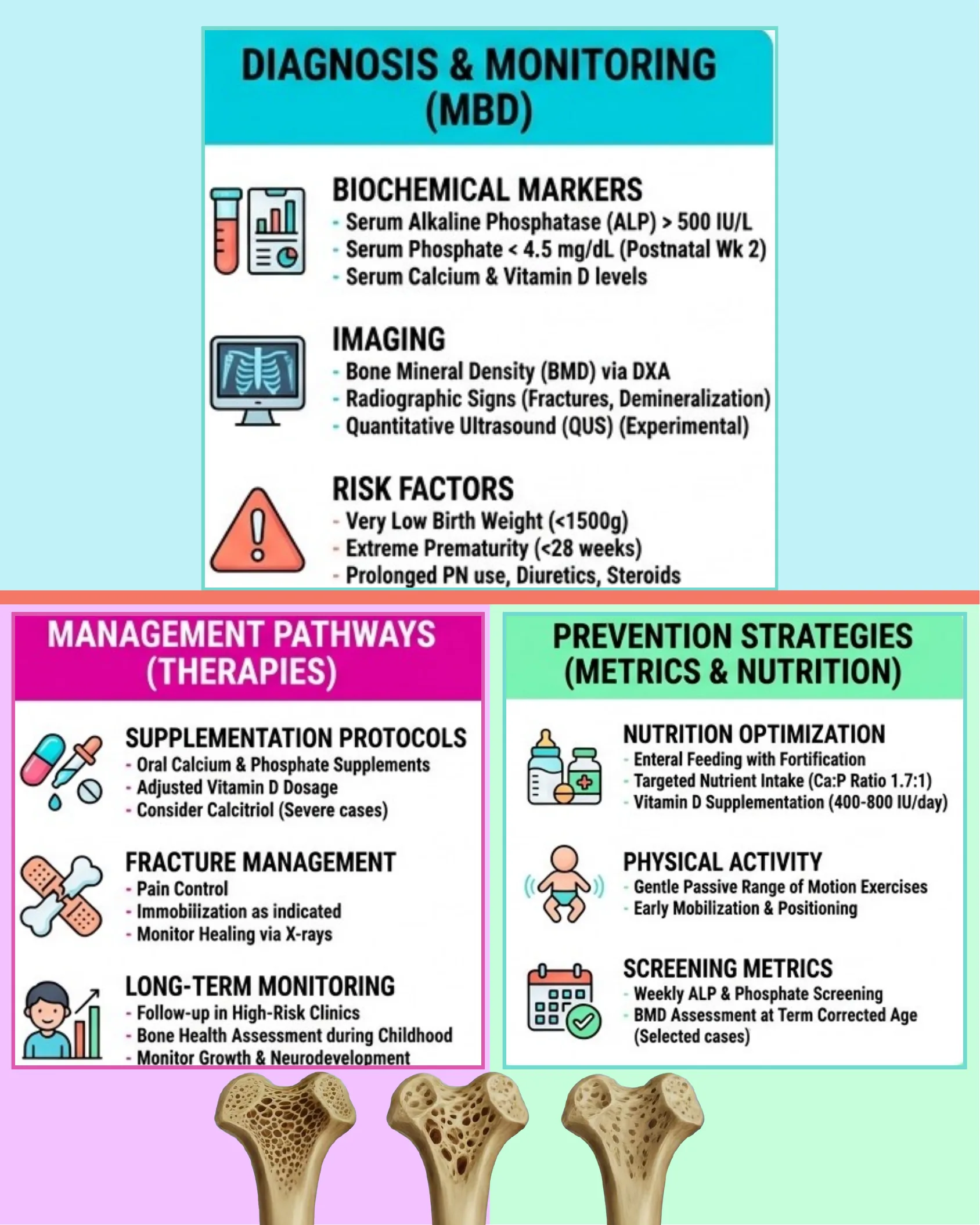
Diagnosis & Monitoring (MBD)
Biochemical Markers
- Serum Alkaline Phosphatase (ALP) > 500 IU/L
- Serum Phosphate < 4.5 mg/dL (Postnatal Wk 2)
- Serum Calcium & Vitamin D levels
Imaging
- Bone Mineral Density (BMD) via DXA
- Radiographic Signs (Fractures, Demineralization)
- Quantitative Ultrasound (QUS) (Experimental)
Risk Factors
- Very Low Birth Weight (<1500g)
- Extreme Prematurity (<28 weeks)
- Prolonged PN use, Diuretics, Steroids
Management Pathways (Therapies)
Supplementation Protocols
- Oral Calcium & Phosphate Supplements
- Adjusted Vitamin D Dosage
- Consider Calcitriol (Severe cases)
Fracture Management
- Pain Control
- Immobilization as indicated
- Monitor Healing via X-rays
Long-Term Monitoring
- Follow-up in High-Risk Clinics
- Bone Health Assessment during Childhood
- Monitor Growth & Neurodevelopment
Prevention Strategies (Metrics & Nutrition)
Nutrition Optimization
- Enteral Feeding with Fortification
- Targeted Nutrient Intake (Ca:P Ratio 1.7:1)
- Vitamin D Supplementation (400-800 IU/day)
Physical Activity
- Gentle Passive Range of Motion Exercises
- Early Mobilization & Positioning
Screening Metrics
- Weekly ALP & Phosphate Screening
- BMD Assessment at Term Corrected Age (Selected cases)
Defining the Mineral Accretion Gap
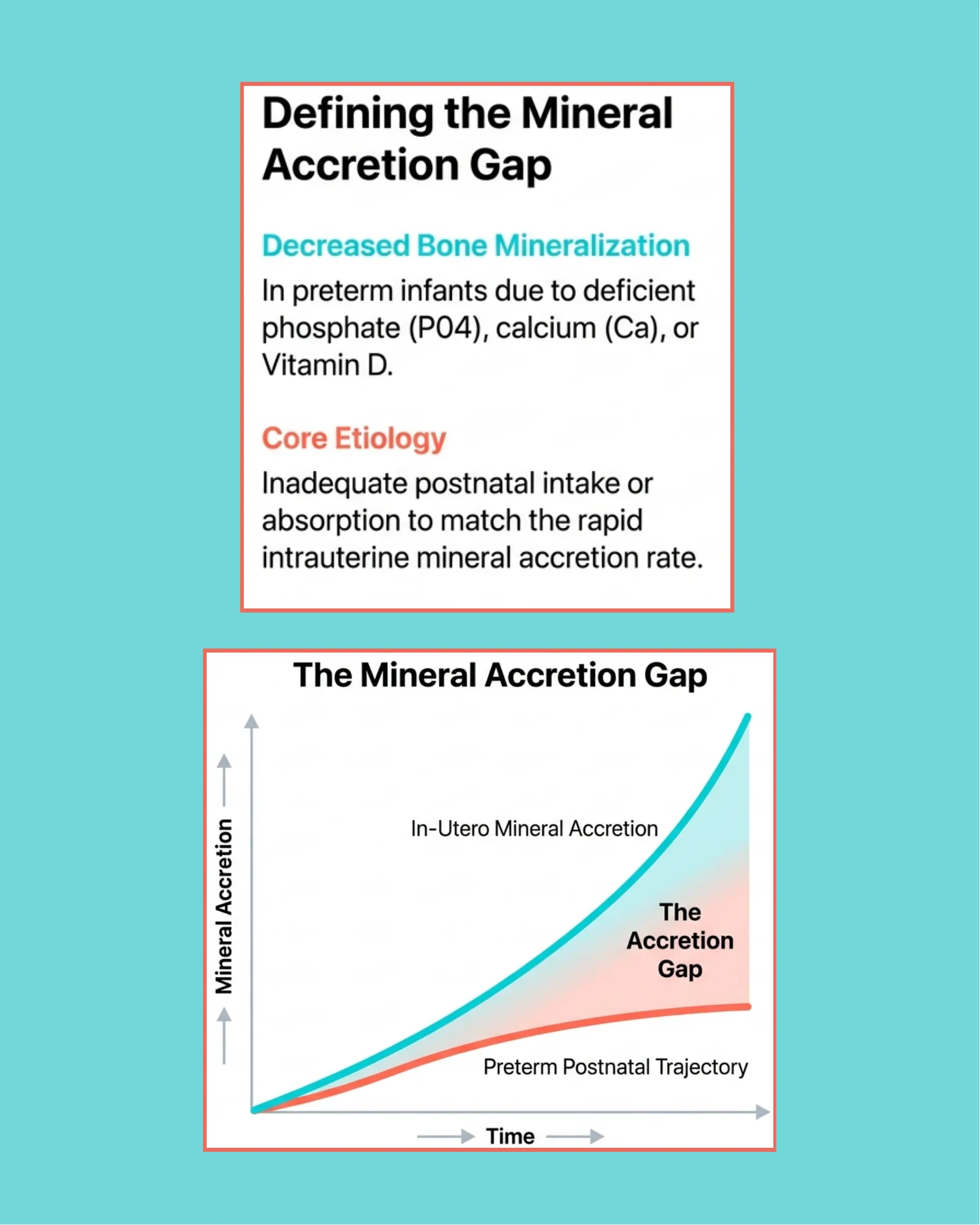
Defining the Mineral Accretion Gap
Decreased Bone Mineralization
In preterm infants due to deficient phosphate (PO4), calcium (Ca), or Vitamin D.
Core Etiology
Inadequate postnatal intake or absorption to match the rapid intrauterine mineral accretion rate.
The Mineral Accretion Gap
Graph showing Mineral Accretion (y-axis) vs Time (x-axis), comparing two curves:
- In-Utero Mineral Accretion (upper curve, rising steeply)
- Preterm Postnatal Trajectory (lower curve, rising slowly)
The widening area between the two curves is labeled The Accretion Gap.
The High-Risk Patient Profile
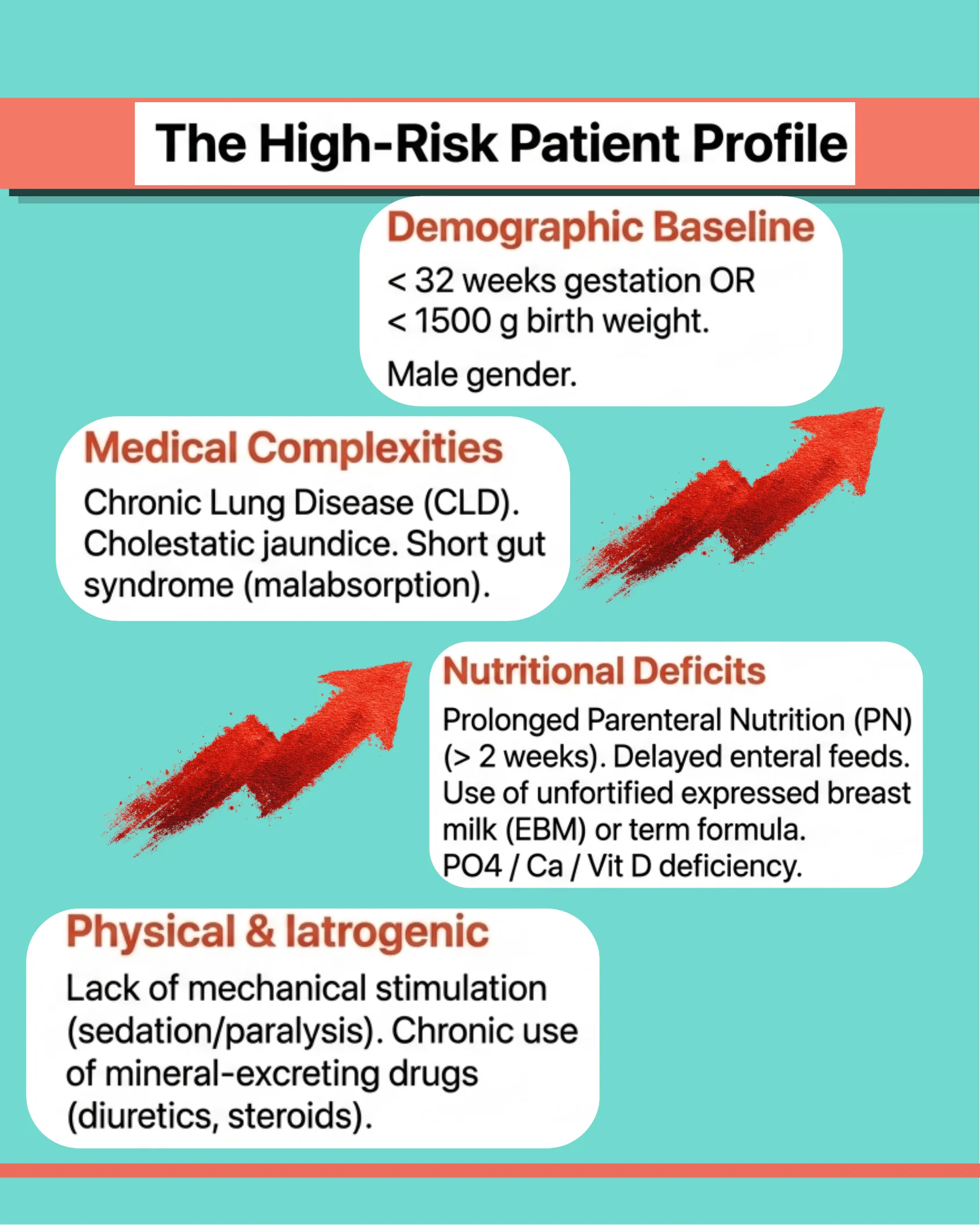
The High-Risk Patient Profile
Demographic Baseline
- < 32 weeks gestation OR < 1500 g birth weight.
- Male gender.
Medical Complexities
- Chronic Lung Disease (CLD).
- Cholestatic jaundice.
- Short gut syndrome (malabsorption).
Nutritional Deficits
- Prolonged Parenteral Nutrition (PN) (> 2 weeks).
- Delayed enteral feeds.
- Use of unfortified expressed breast milk (EBM) or term formula.
- PO4 / Ca / Vit D deficiency.
Physical & Iatrogenic
- Lack of mechanical stimulation (sedation/paralysis).
- Chronic use of mineral-excreting drugs (diuretics, steroids).
The Silent Progression: Clinical Timeline
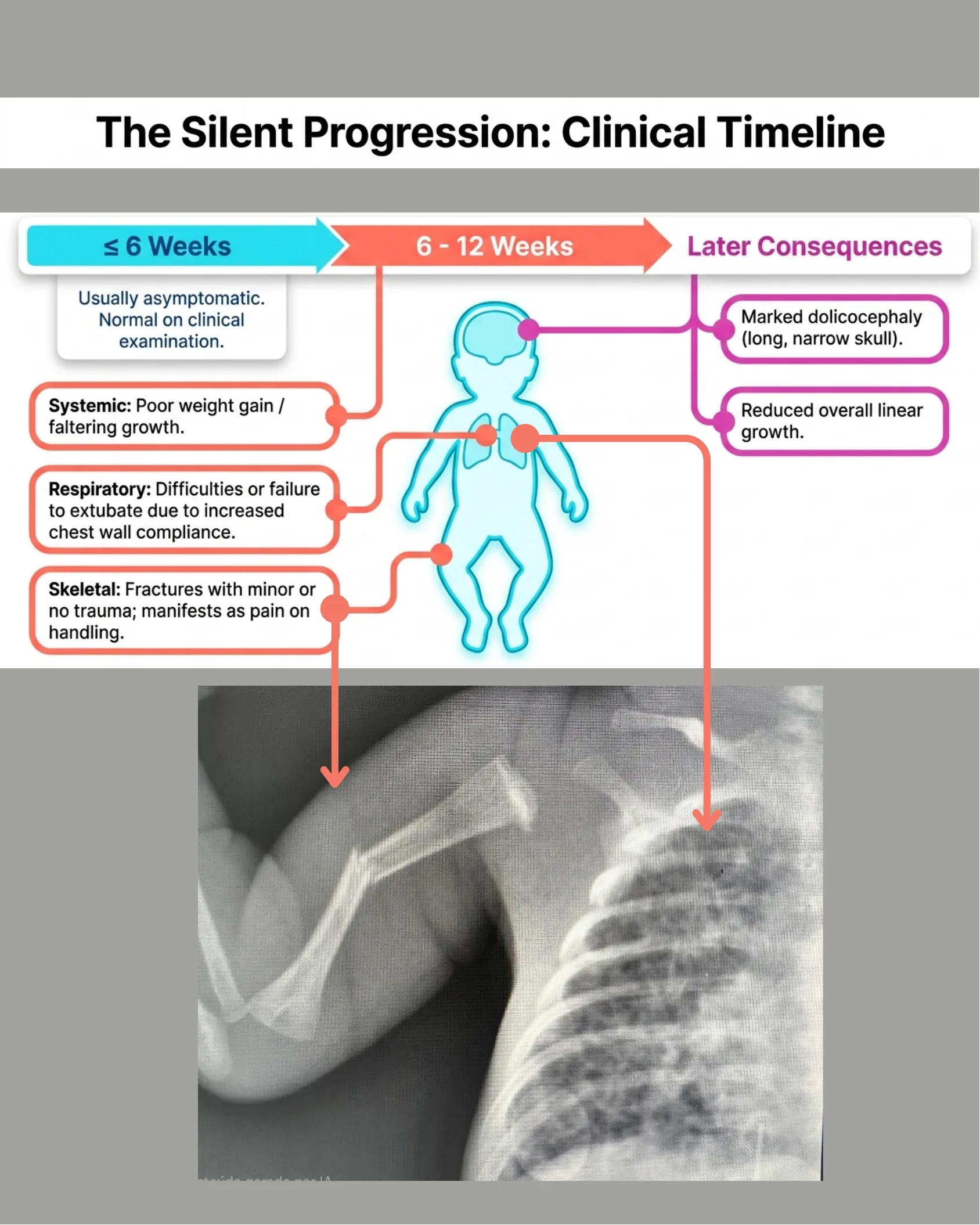
The Silent Progression: Clinical Timeline
≤ 6 Weeks
Usually asymptomatic. Normal on clinical examination.
6 – 12 Weeks
- Systemic: Poor weight gain / faltering growth.
- Respiratory: Difficulties or failure to extubate due to increased chest wall compliance.
- Skeletal: Fractures with minor or no trauma; manifests as pain on handling.
Later Consequences
- Marked dolicocephaly (long, narrow skull).
- Reduced overall linear growth.
Diagnostic Biomarkers & Radiology
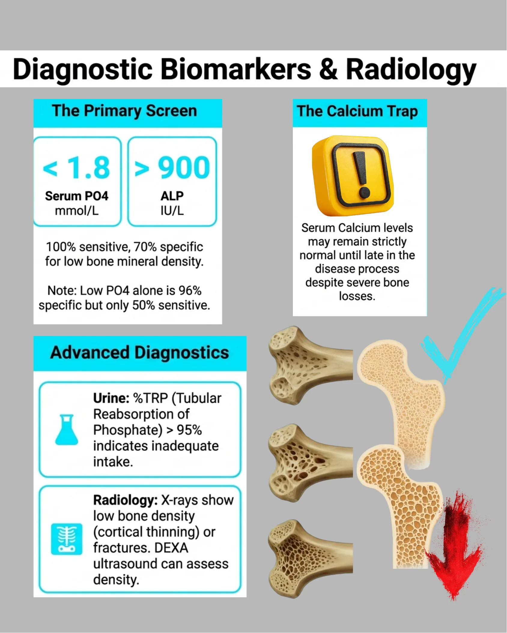
Diagnostic Biomarkers & Radiology
The Primary Screen
- < 1.8 mmol/L Serum PO4
- > 900 IU/L ALP
100% sensitive, 70% specific for low bone mineral density.
Note: Low PO4 alone is 96% specific but only 50% sensitive.
The Calcium Trap
Serum Calcium levels may remain strictly normal until late in the disease process despite severe bone losses.
Advanced Diagnostics
- Urine: %TRP (Tubular Reabsorption of Phosphate) > 95% indicates inadequate intake.
- Radiology: X-rays show low bone density (cortical thinning) or fractures. DEXA ultrasound can assess density.
The Three Pillars of Prevention
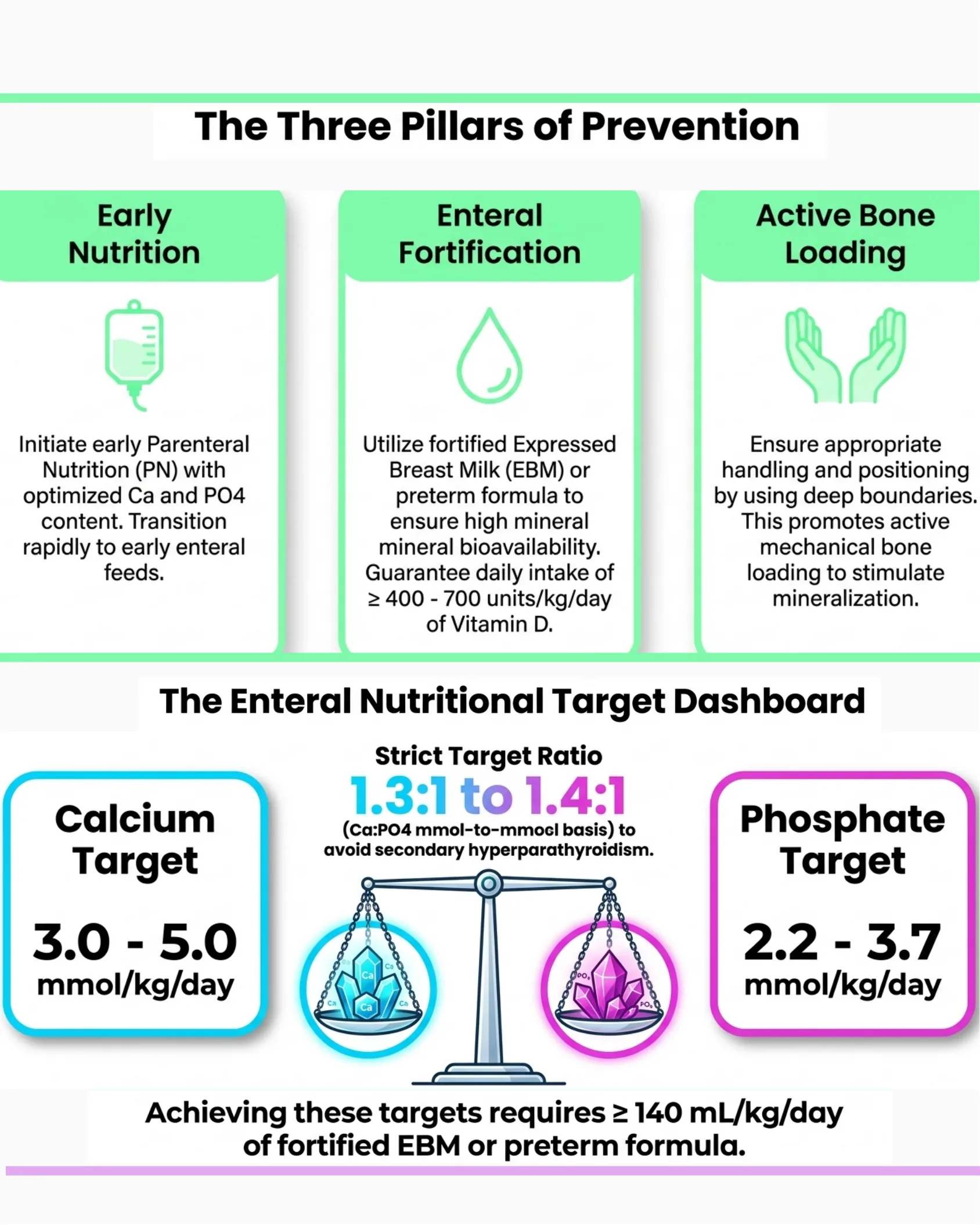
The Three Pillars of Prevention
Early Nutrition
Initiate early Parenteral Nutrition (PN) with optimized Ca and PO4 content. Transition rapidly to early enteral feeds.
Enteral Fortification
Utilize fortified Expressed Breast Milk (EBM) or preterm formula to ensure high mineral bioavailability. Guarantee daily intake of ≥ 400 – 700 units/kg/day of Vitamin D.
Active Bone Loading
Ensure appropriate handling and positioning by using deep boundaries. This promotes active mechanical bone loading to stimulate mineralization.
The Enteral Nutritional Target Dashboard
- Calcium Target: 3.0 – 5.0 mmol/kg/day
- Phosphate Target: 2.2 – 3.7 mmol/kg/day
- Strict Target Ratio: 1.3:1 to 1.4:1 (Ca:PO4 mmol-to-mmol basis) to avoid secondary hyperparathyroidism.
Achieving these targets requires ≥ 140 mL/kg/day of fortified EBM or preterm formula.
The Week 3 Screening Trigger & PTH Diagnostic Matrix
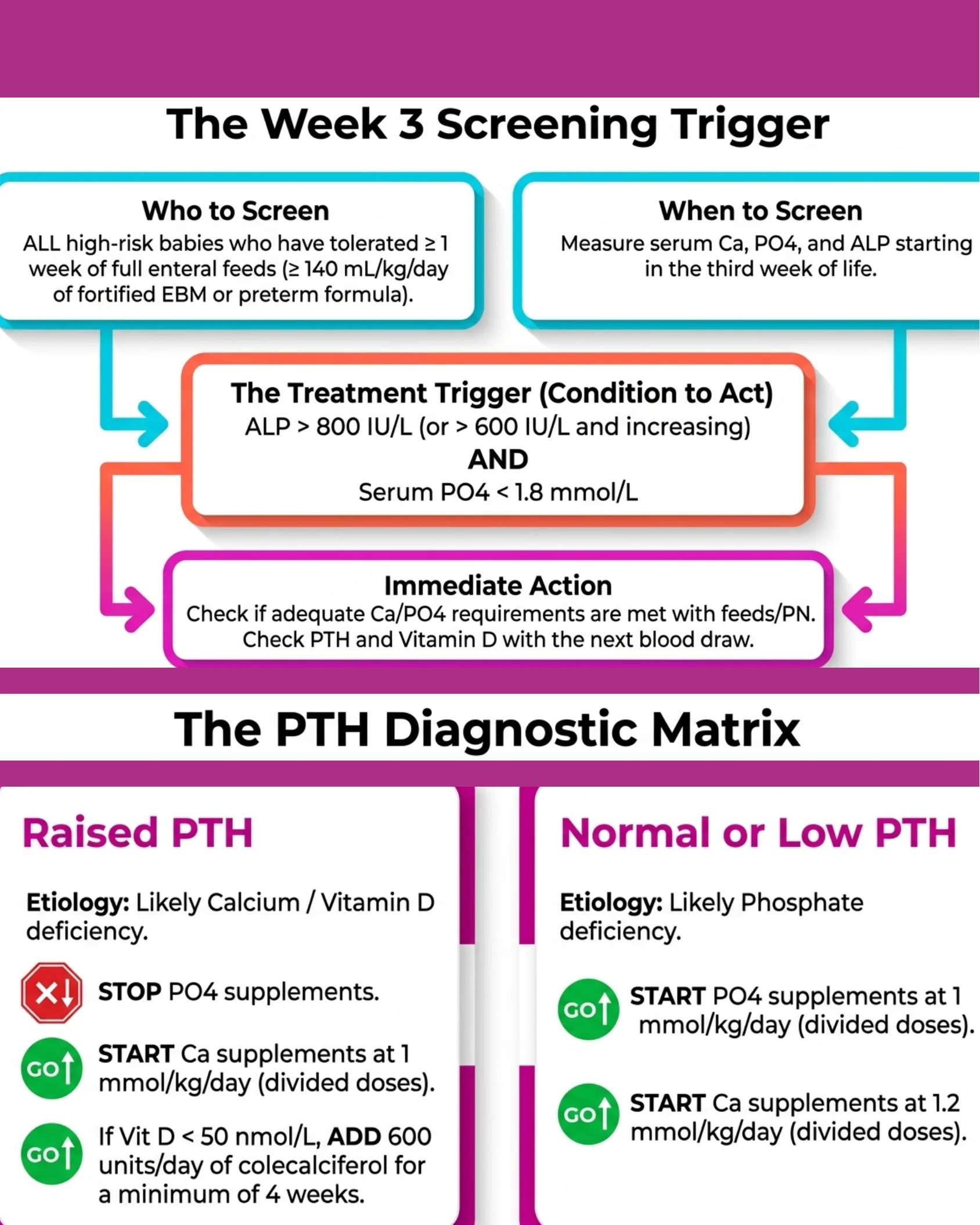
The Week 3 Screening Trigger
Who to Screen
ALL high-risk babies who have tolerated ≥ 1 week of full enteral feeds (≥ 140 mL/kg/day of fortified EBM or preterm formula).
When to Screen
Measure serum Ca, PO4, and ALP starting in the third week of life.
The Treatment Trigger (Condition to Act)
ALP > 800 IU/L (or > 600 IU/L and increasing) AND Serum PO4 < 1.8 mmol/L
Immediate Action
Check if adequate Ca/PO4 requirements are met with feeds/PN. Check PTH and Vitamin D with the next blood draw.
The PTH Diagnostic Matrix
Raised PTH
Etiology: Likely Calcium / Vitamin D deficiency.
- STOP PO4 supplements.
- START Ca supplements at 1 mmol/kg/day (divided doses).
- If Vit D < 50 nmol/L, ADD 600 units/day of colecalciferol for a minimum of 4 weeks.
Normal or Low PTH
Etiology: Likely Phosphate deficiency.
- START PO4 supplements at 1 mmol/kg/day (divided doses).
- START Ca supplements at 1.2 mmol/kg/day (divided doses).
Crucial Administration Warnings (The Do Nots) & Ongoing Monitoring and Resolution
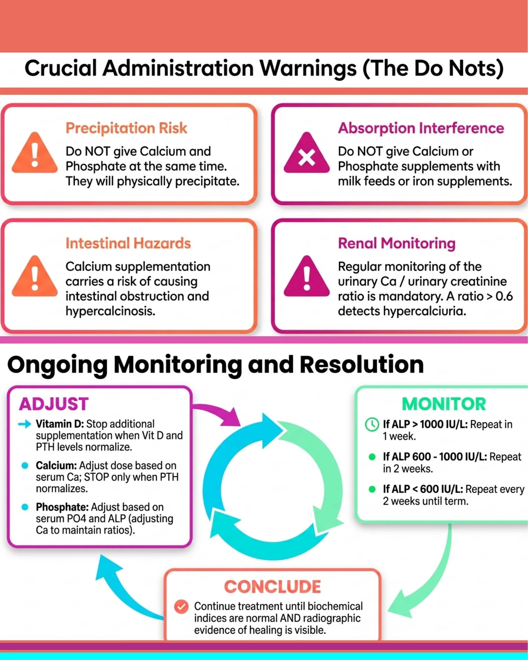
Crucial Administration Warnings (The Do Nots)
- Precipitation Risk: Do NOT give Calcium and Phosphate at the same time. They will physically precipitate.
- Absorption Interference: Do NOT give Calcium or Phosphate supplements with milk feeds or iron supplements.
- Intestinal Hazards: Calcium supplementation carries a risk of causing intestinal obstruction and hypercalcinosis.
- Renal Monitoring: Regular monitoring of the urinary Ca / urinary creatinine ratio is mandatory. A ratio > 0.6 detects hypercalciuria.
Ongoing Monitoring and Resolution
- MONITOR:
- If ALP > 1000 IU/L: Repeat in 1 week.
- If ALP 600 – 1000 IU/L: Repeat in 2 weeks.
- If ALP < 600 IU/L: Repeat every 2 weeks until term.
- ADJUST:
- Vitamin D: Stop additional supplementation when Vit D and PTH levels normalize.
- Calcium: Adjust dose based on serum Ca; STOP only when PTH normalizes.
- Phosphate: Adjust based on serum PO4 and ALP (adjusting Ca to maintain ratios).
- CONCLUDE: Continue treatment until biochemical indices are normal AND radiographic evidence of healing is visible.
Neofast – Neonatal Prescription

Neofast
Neonatal Prescription
- Download on the App Store
- Get it on Google Play
Frequently asked questions
How is metabolic bone disease of prematurity diagnosed?
By biochemical markers — ALP above 500 IU/L and phosphate below 4.5 mg/dL (postnatal week 2) — supported by imaging such as DXA or radiographs.
Which preterm infants are at highest risk?
Those below 32 weeks or under 1500 g, especially with chronic lung disease, cholestasis, short gut, or parenteral nutrition beyond 2 weeks.
What Ca:P ratio and vitamin D dose are recommended?
A calcium-to-phosphate ratio of 1.7:1 with vitamin D 400–800 IU/day.