Key takeaways
- Antenatal corticosteroids given as a complete course before 34 weeks reduce IVH incidence by more than 50% by accelerating germinal matrix vessel maturation.
- Over 90% of all GMH-IVH cases manifest within the first 3 days of life, making Day 1 and Day 3 cranial ultrasound scans critical for detection.
- The PHVD intervention action line is Ventricular Index (VI) greater than 97th percentile + 4 mm AND Anterior Horn Width (AHW) greater than 6 mm, and therapy should start before clinical signs of raised intracranial pressure appear.
- Grade III IVH carries a 15-25% cerebral palsy risk and 30-50% of these infants develop Post-Hemorrhagic Ventricular Dilatation (PHVD), which raises CP risk to 30-40%.
- Periventricular Hemorrhagic Infarction (PVHI) carries roughly 50% risk of unilateral spastic hemiplegia, with non-myelination of the PLIC on term-equivalent age MRI as the primary predictor.
References
- Primary Source: Authors. Neonatal Brain Injury: An Illustrated Clinical Guide. Open Access Perinatal & Neonatal Neurology Series.
- Volpe JJ. Volpe’s Neurology of the Newborn. 6th ed. Philadelphia, PA: Elsevier; 2018. Chapter 24: Germinal Matrix-Intraventricular Hemorrhage of the Premature Infant.
- Levene MI. Measurement of the growth of the lateral ventricles in preterm infants with posthaemorrhagic ventricular dilatation. Arch Dis Child. 1981;56(12):900-904.
- Whitelaw A, Aquilina K. Posthaemorrhagic ventricular dilatation in the premature infant: natural history and management. Arch Dis Child Fetal Neonatal Ed. 2012;97(5):F384-F388.
- De Vries LS, Groenendaal F. Neuroimaging in the preterm infant. Ment Retard Dev Disabil Res Rev. 2002;8(1):3-12.
- ELFIN & DRIFT Trial Investigators. Randomised trial of DRIFT (drain, irrigate and forecast cure) in posthaemorrhagic ventricular dilatation. Pediatrics. 2010;125(5):e1079-e1085.
- Brouwer MJ, de Vries LS, Pistorius LR, et al. Posthemorrhagic ventricular dilatation: a comparison of two intervention thresholds. Neonatology. 2013;104(1):49-55.
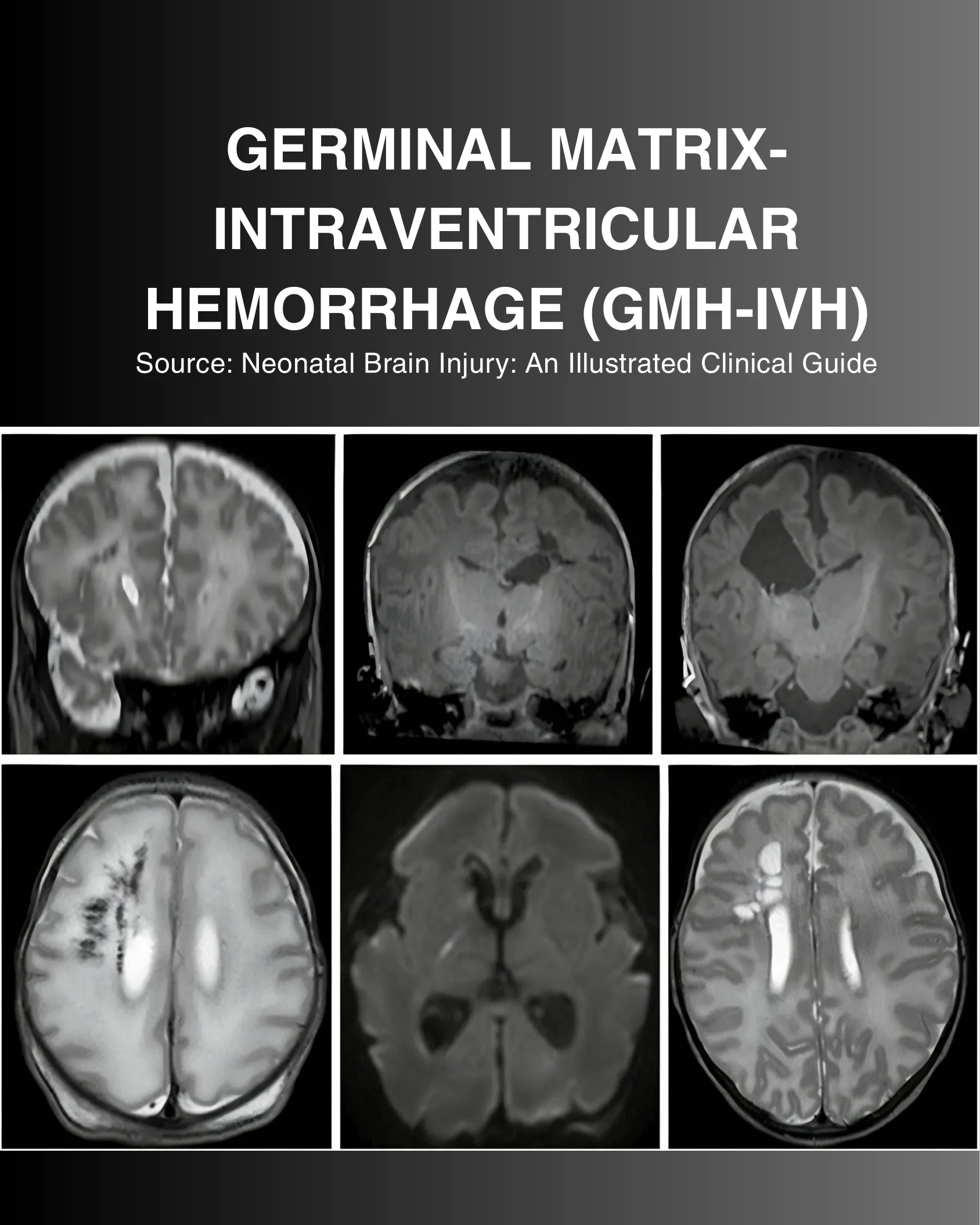
GERMINAL MATRIX-INTRAVENTRICULAR HEMORRHAGE (GMH-IVH)
Source: Neonatal Brain Injury: An Illustrated Clinical Guide
1. Etiology, Anatomy and Pathophysiologic Vulnerability
Germinal Matrix-Intraventricular Hemorrhage (GMH-IVH) remains one of the most critical neurological complications in extremely preterm infants (<32 weeks gestation, especially <28 weeks). The primary anatomical origin is the germinal matrix: a highly vascularized, cellular subependymal structure located along the lateral ventricles that serves as the microenvironment for neuronal and glial progenitor proliferation.
Key Vascular Vulnerability Factors:
- Immature Vascular Architecture: The capillaries of the germinal matrix consist of a single layer of endothelial cells with minimal basement membrane support and sparse pericytes or astrocytic end-feet.
- Impaired Cerebral Autoregulation: Extremely preterm neonates frequently exhibit a ‘pressure-passive’ cerebral circulation, where systemic arterial blood pressure fluctuations directly transmit to fragile germinal matrix microvessels.
- High Metabolic & Blood Flow Demand: The rapid cellular turnover in the subependymal zone requires high regional perfusion, rendering it hyperemic and vulnerable to rupture during pressure surges.
- Fibrinolytic Activity & Venous Stasis: High tissue plasminogen activator levels within the germinal matrix inhibit clot stability, while deep medullary venous congestion accelerates vessel rupture.
2. Volpe Classification and Parenchymal Involvement
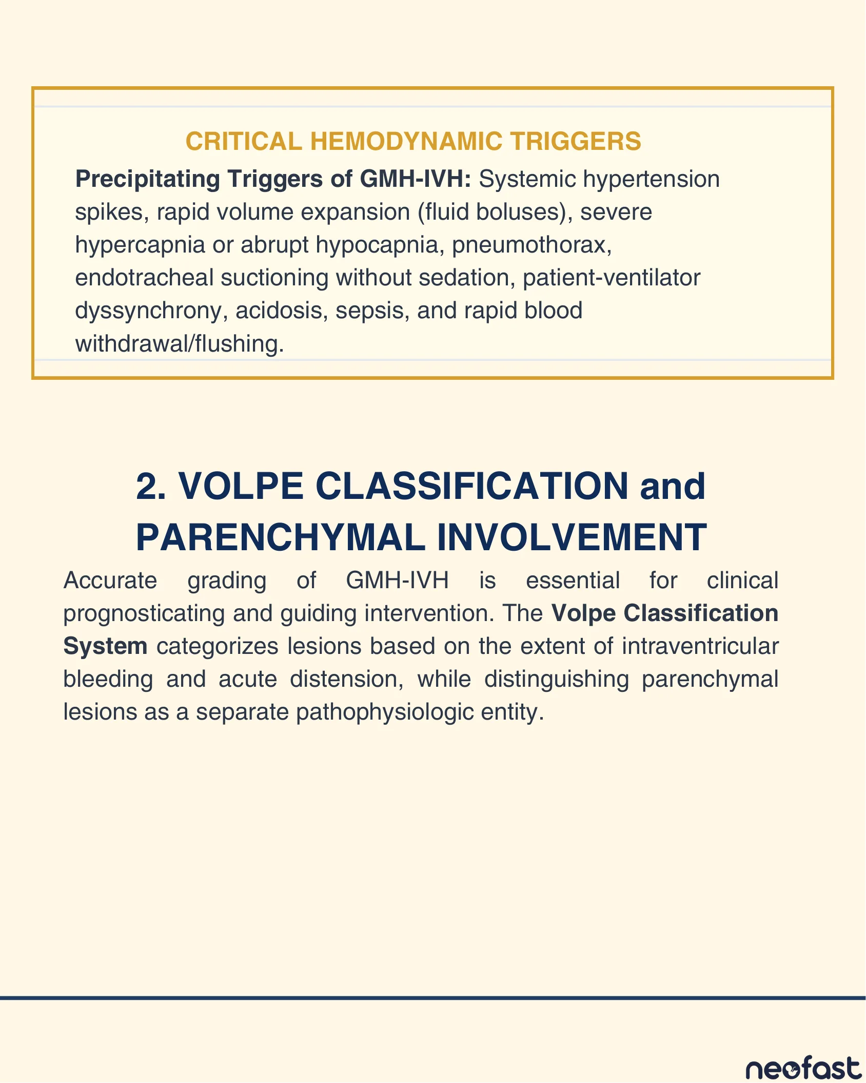
Critical Hemodynamic Triggers
Precipitating Triggers of GMH-IVH: Systemic hypertension spikes, rapid volume expansion (fluid boluses), severe hypercapnia or abrupt hypocapnia, pneumothorax, endotracheal suctioning without sedation, patient-ventilator dyssynchrony, acidosis, sepsis, and rapid blood withdrawal/flushing.
2. Volpe Classification and Parenchymal Involvement
Accurate grading of GMH-IVH is essential for clinical prognosticating and guiding intervention. The Volpe Classification System categorizes lesions based on the extent of intraventricular bleeding and acute distension, while distinguishing parenchymal lesions as a separate pathophysiologic entity.
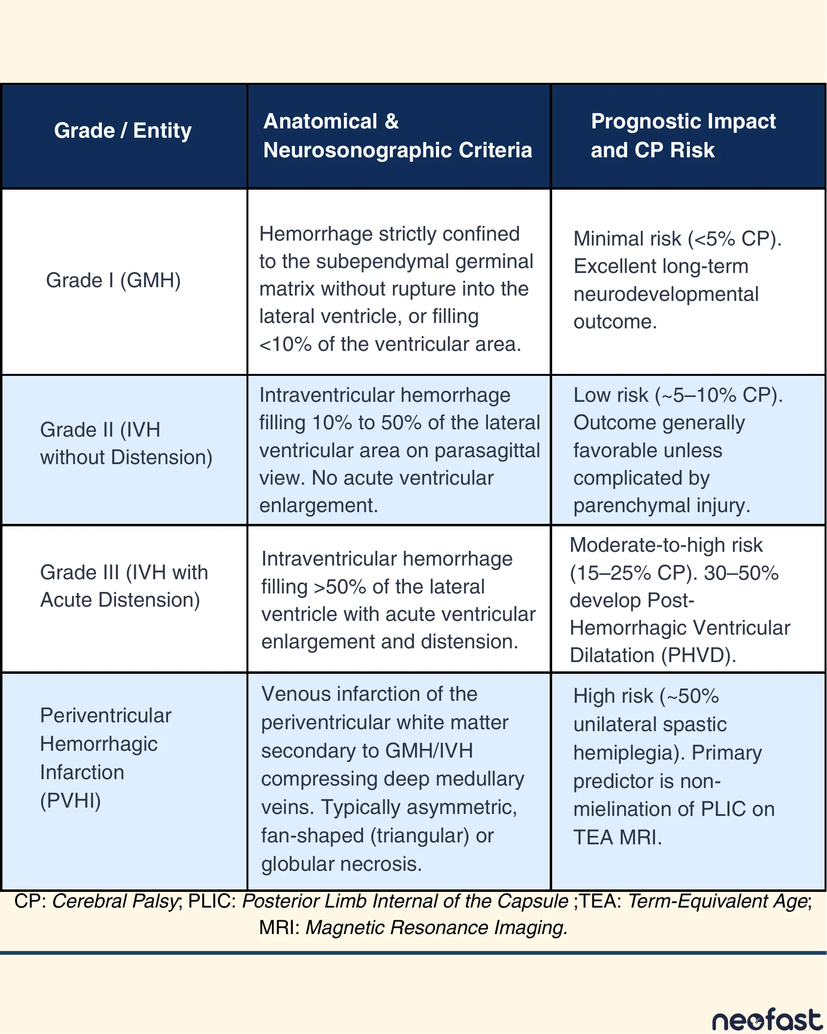
Volpe Grading Table
| Grade / Entity | Anatomical & Neurosonographic Criteria | Prognostic Impact and CP Risk |
|---|---|---|
| Grade I (GMH) | Hemorrhage strictly confined to the subependymal germinal matrix without rupture into the lateral ventricle, or filling <10% of the ventricular area. | Minimal risk (<5% CP). Excellent long-term neurodevelopmental outcome. |
| Grade II (IVH without Distension) | Intraventricular hemorrhage filling 10% to 50% of the lateral ventricular area on parasagittal view. No acute ventricular enlargement. | Low risk (~5–10% CP). Outcome generally favorable unless complicated by parenchymal injury. |
| Grade III (IVH with Acute Distension) | Intraventricular hemorrhage filling >50% of the lateral ventricle with acute ventricular enlargement and distension. | Moderate-to-high risk (15–25% CP). 30–50% develop Post-Hemorrhagic Ventricular Dilatation (PHVD). |
| Periventricular Hemorrhagic Infarction (PVHI) | Venous infarction of the periventricular white matter secondary to GMH/IVH compressing deep medullary veins. Typically asymmetric, fan-shaped (triangular) or globular necrosis. | High risk (~50% unilateral spastic hemiplegia). Primary predictor is non-mielination of PLIC on TEA MRI. |
CP: Cerebral Palsy; PLIC: Posterior Limb Internal of the Capsule; TEA: Term-Equivalent Age; MRI: Magnetic Resonance Imaging.
3. Primary Prevention Strategies: Antenatal & NICU Care Bundles
Because established IVH has no direct reversal agent, prevention is the cornerstone of clinical management. Evidence-based prevention requires an integrated approach combining antenatal maternal therapy with strict postnatal hemodynamic protection.
A. Antenatal Interventions:
- Antenatal Corticosteroids (ANS): Administration of complete course betamethasone/dexamethasone to mothers at risk of preterm delivery <34 weeks reduces IVH incidence by >50% by accelerating vessel maturation in the germinal matrix.
- Magnesium Sulfate (MgSO4): Antenatal neuroprotective infusion in women presenting with imminently expected preterm labor (<32 weeks) stabilizes neuronal cell membranes and microvascular tone.
- In Utero Transfer: Maternal transfer to a high-volume tertiary perinatal center to avoid out-of-hospital birth and traumatic transport.
3. Primary Prevention Strategies: NICU Neuroprotective Care Bundle
B. Postnatal ‘NICU Neuroprotective Care Bundle’ (First 72–96 Hours):
- Head Positioning: Maintain the infant’s head strictly in the neutral midline position with the head of the bed elevated 15°. Prevents jugular venous obstruction and fluctuations in intracranial venous pressure.
- Minimal Handling & Clustered Care: Group nursing procedures to avoid frequent disturbances. Avoid sudden noxious stimuli, loud noises, or unnecessary physical rotation.
- Gentle Diaper Changes: Do NOT elevate the infant’s legs above head level during diaper changes (which causes a sudden surge in central venous pressure). Instead, gently roll the infant laterally.
- Cautious Fluid Administration: Avoid routine rapid boluses of crystalloids or colloid solutions. Infuse all intravenous drugs and blood products slowly (over ≥30–60 minutes).
- Cateter & Blood Sampling Protocols: Avoid rapid withdrawal or forceful flushing of umbilical arterial/venous catheters.
- Targeted Mechanical Ventilation: Maintain strict normocapnia (PaCO2 35–45 mmHg). Avoid acute hypercapnia (which causes profound cerebral vasodilation) and severe hypocapnia (which induces vasoconstriction and ischemic injury).
4. Neuroimaging Protocols & Serial Ultrasound Monitoring
Cranial Ultrasound (cUS) is the primary bedside screening and monitoring tool for GMH-IVH due to its accessibility, absence of radiation, and high sensitivity for hemorrhage and ventricular distension. Magnetic Resonance Imaging (MRI) serves as a problem-solving and long-term prognosticating tool.
Recommended cUS Screening Timeline:
- Day 1 (0–24 Hours): Baseline scan to detect early-onset IVH (16% occur in first hour; 50% by 6 hours).
- Day 3 (48–72 Hours): Critical scan—over 90% of all GMH-IVH cases manifest within the first 3 days of life.
- Day 7 & Day 14: Screening for delayed-onset hemorrhage, early ventricular enlargement, or white matter flares.
- Term Equivalent Age (TEA): Final structured ultrasound to assess ventriculomegaly, porencephaly, and cystic leukomalacia.
- Serial Monitoring Protocol for Grade III/PHVD: Perform cUS 2–3 times weekly (or daily during active LP/reservoir drainage) to plot Ventricular Index (VI) and Anterior Horn Width (AHW).
Role of Advanced MRI
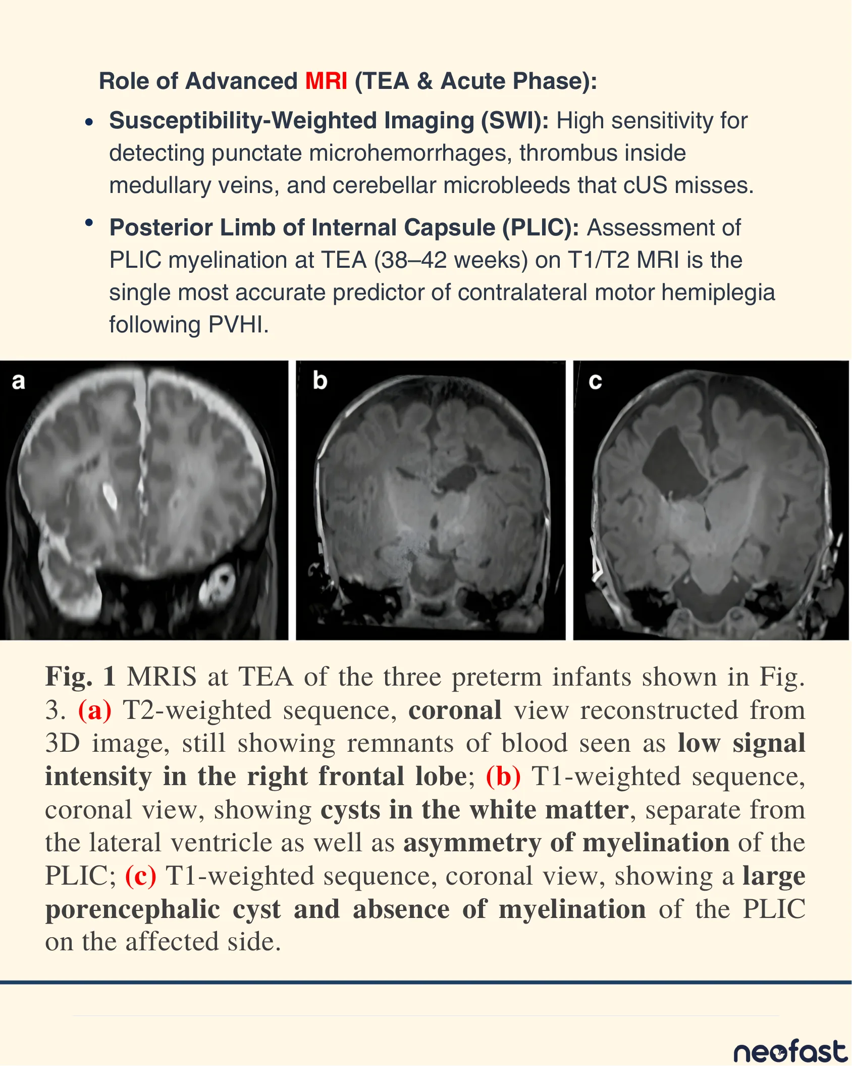
Role of Advanced MRI (TEA & Acute Phase):
- Susceptibility-Weighted Imaging (SWI): High sensitivity for detecting punctate microhemorrhages, thrombus inside medullary veins, and cerebellar microbleeds that cUS misses.
- Posterior Limb of Internal Capsule (PLIC): Assessment of PLIC myelination at TEA (38–42 weeks) on T1/T2 MRI is the single most accurate predictor of contralateral motor hemiplegia following PVHI.
Fig. 1 MRIS at TEA of the three preterm infants shown in Fig. 3. (a) T2-weighted sequence, coronal view reconstructed from 3D image, still showing remnants of blood seen as low signal intensity in the right frontal lobe; (b) T1-weighted sequence, coronal view, showing cysts in the white matter, separate from the lateral ventricle as well as asymmetry of myelination of the PLIC; (c) T1-weighted sequence, coronal view, showing a large porencephalic cyst and absence of myelination of the PLIC on the affected side.
Fig. 2 — Brain MRI of PVHI in a Preterm Infant
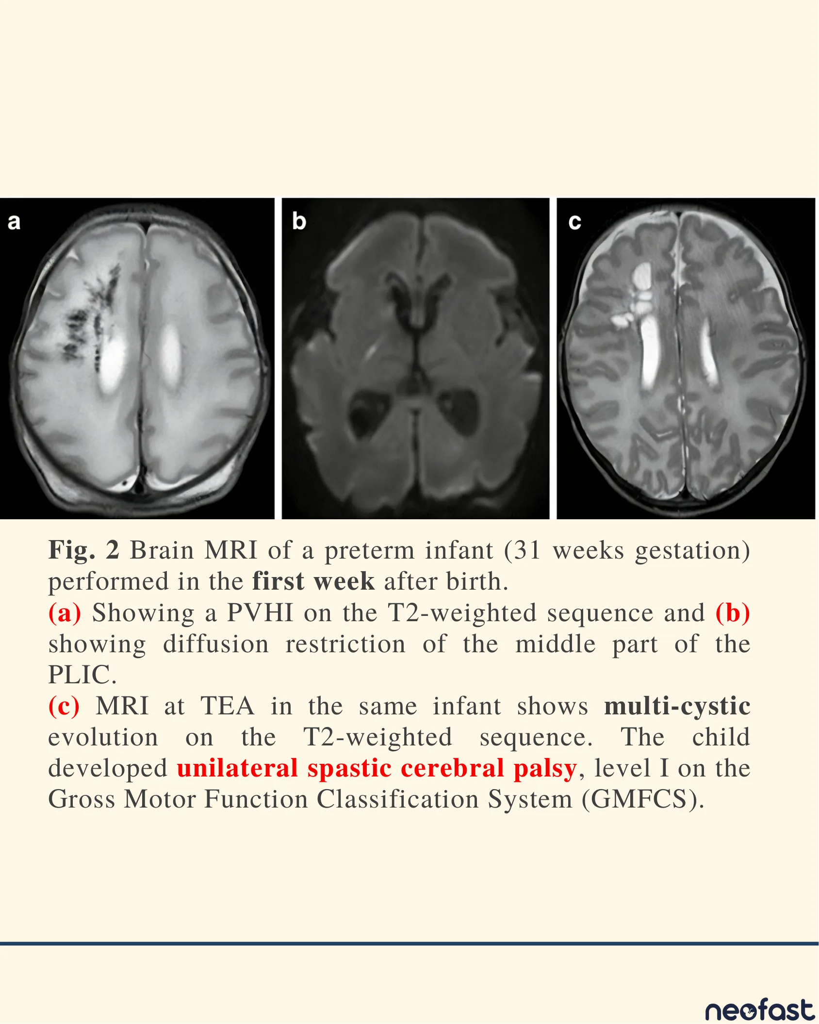
Fig. 2 Brain MRI of a preterm infant (31 weeks gestation) performed in the first week after birth.
(a) Showing a PVHI on the T2-weighted sequence and (b) showing diffusion restriction of the middle part of the PLIC.
(c) MRI at TEA in the same infant shows multi-cystic evolution on the T2-weighted sequence. The child developed unilateral spastic cerebral palsy, level I on the Gross Motor Function Classification System (GMFCS).
Figure 3: Cranial Ultrasound (Coronal & Parasagittal Views)
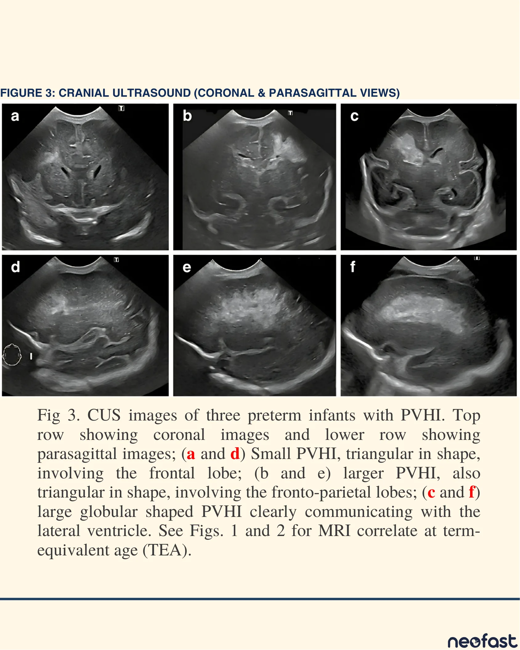
Figure 3: Cranial Ultrasound (Coronal & Parasagittal Views)
Fig 3. CUS images of three preterm infants with PVHI. Top row showing coronal images and lower row showing parasagittal images; (a and d) Small PVHI, triangular in shape, involving the frontal lobe; (b and e) larger PVHI, also triangular in shape, involving the fronto-parietal lobes; (c and f) large globular shaped PVHI clearly communicating with the lateral ventricle. See Figs. 1 and 2 for MRI correlate at term-equivalent age (TEA).
Figure 4: Cranial Ultrasound (Coronal & Parasagittal Views)
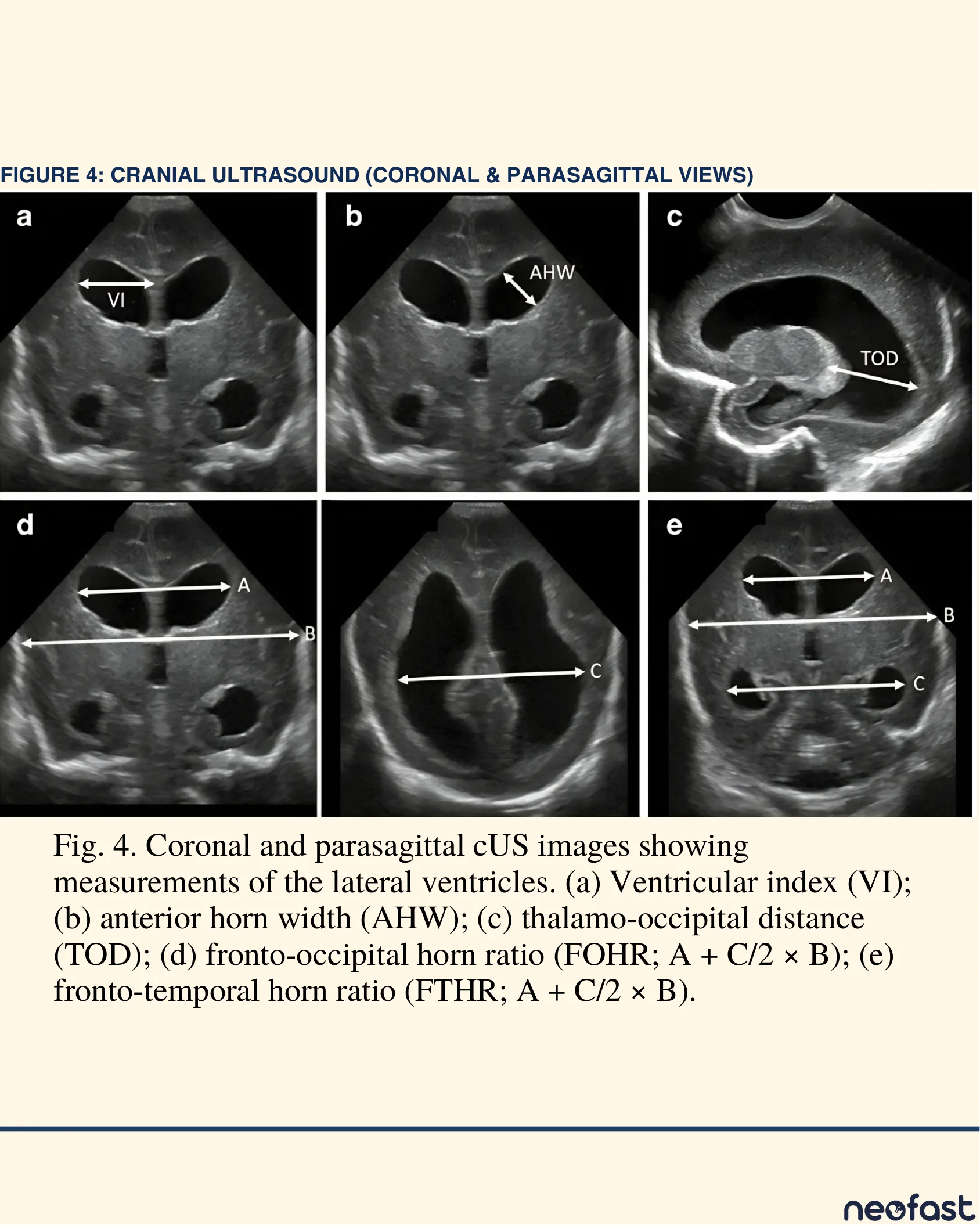
Figure 4: Cranial Ultrasound (Coronal & Parasagittal Views)
Fig. 4. Coronal and parasagittal cUS images showing measurements of the lateral ventricles. (a) Ventricular index (VI); (b) anterior horn width (AHW); (c) thalamo-occipital distance (TOD); (d) fronto-occipital horn ratio (FOHR; A + C/2 × B); (e) fronto-temporal horn ratio (FTHR; A + C/2 × B).
5. Post-Hemorrhagic Ventricular Dilatation (PHVD) Management
5. Post-Hemorrhagic Ventricular Dilatation (PHVD) Management
PHVD develops in 30–50% of infants with Grade III IVH due to obliterative arachnoiditis in the posterior fossa or aqueductal obstruction. Early intervention based on objective neurosonographic thresholds prevents progressive white matter compression, axonal stretch, and ischemic damage.
Quantitative Ultrasound Metrics:
- Ventricular Index (VI – Levene): Distance from falce to lateral wall of anterior horn in coronal view (at foramen of Monro).
- Anterior Horn Width (AHW): Diagonal measure of the anterior horn at its widest point in coronal view.
- Thalamo-Occipital Distance (TOD): Distance from thalamus to posterior wall of occipital horn in parasagittal view.
Critical Action Threshold for Intervention
PHVD Intervention Threshold:
- Action Line for Early Intervention: Initiate therapy when Ventricular Index (VI) > 97th percentile + 4 mm AND Anterior Horn Width (AHW) > 6 mm.
- Do NOT wait for clinical signs of raised intracranial pressure (e.g., bulging fontanelle, sun-setting eyes, rapid head circumference acceleration), as irreversible white matter distortion occurs prior to clinical signs.
Sequential Intervention Algorithm
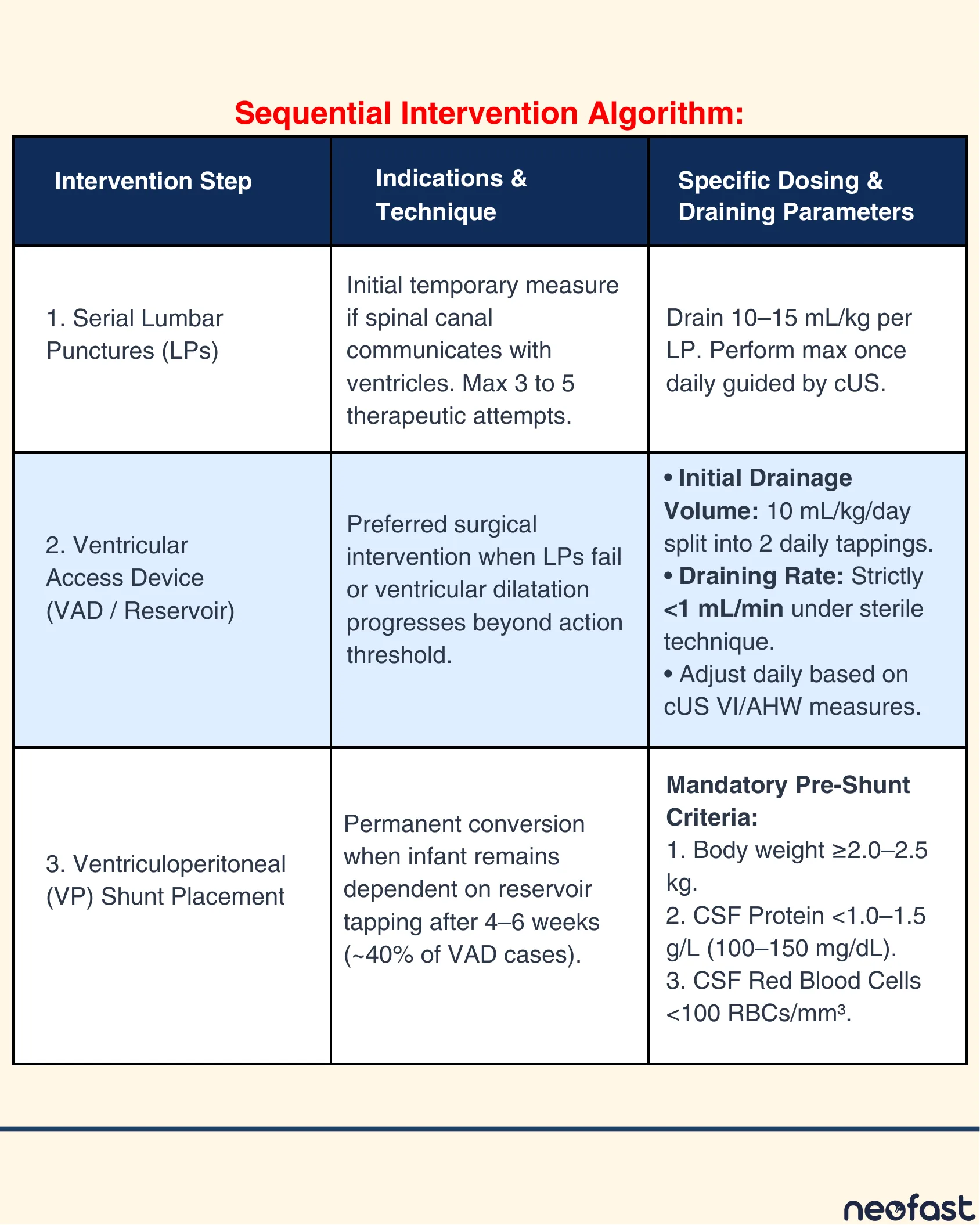
Sequential Intervention Algorithm:
| Intervention Step | Indications & Technique | Specific Dosing & Draining Parameters |
|---|---|---|
| 1. Serial Lumbar Punctures (LPs) | Initial temporary measure if spinal canal communicates with ventricles. Max 3 to 5 therapeutic attempts. | Drain 10–15 mL/kg per LP. Perform max once daily guided by cUS. |
| 2. Ventricular Access Device (VAD / Reservoir) | Preferred surgical intervention when LPs fail or ventricular dilatation progresses beyond action threshold. | Initial Drainage Volume: 10 mL/kg/day split into 2 daily tappings. Draining Rate: Strictly <1 mL/min under sterile technique. Adjust daily based on cUS VI/AHW measures. |
| 3. Ventriculoperitoneal (VP) Shunt Placement | Permanent conversion when infant remains dependent on reservoir tapping after 4–6 weeks (~40% of VAD cases). | Mandatory Pre-Shunt Criteria: 1. Body weight ≥2.0–2.5 kg. 2. CSF Protein <1.0–1.5 g/L (100–150 mg/dL). 3. CSF Red Blood Cells <100 RBCs/mm³. |
6. Neurodevelopmental Outcomes & Long-Term Prognosis
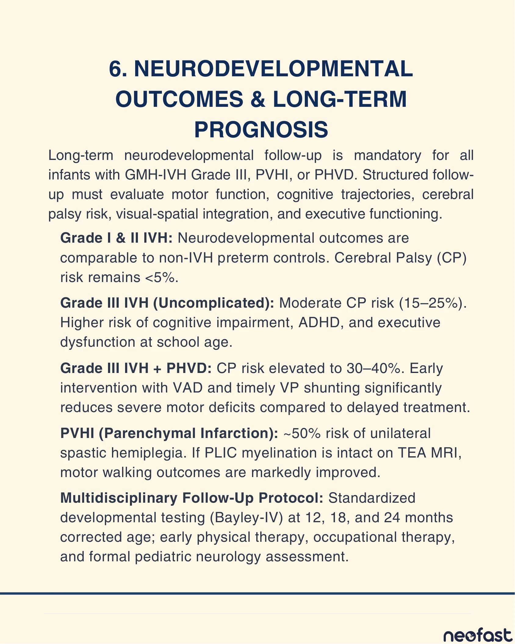
6. Neurodevelopmental Outcomes & Long-Term Prognosis
Long-term neurodevelopmental follow-up is mandatory for all infants with GMH-IVH Grade III, PVHI, or PHVD. Structured follow-up must evaluate motor function, cognitive trajectories, cerebral palsy risk, visual-spatial integration, and executive functioning.
- Grade I & II IVH: Neurodevelopmental outcomes are comparable to non-IVH preterm controls. Cerebral Palsy (CP) risk remains <5%.
- Grade III IVH (Uncomplicated): Moderate CP risk (15–25%). Higher risk of cognitive impairment, ADHD, and executive dysfunction at school age.
- Grade III IVH + PHVD: CP risk elevated to 30–40%. Early intervention with VAD and timely VP shunting significantly reduces severe motor deficits compared to delayed treatment.
- PVHI (Parenchymal Infarction): ~50% risk of unilateral spastic hemiplegia. If PLIC myelination is intact on TEA MRI, motor walking outcomes are markedly improved.
- Multidisciplinary Follow-Up Protocol: Standardized developmental testing (Bayley-IV) at 12, 18, and 24 months corrected age; early physical therapy, occupational therapy, and formal pediatric neurology assessment.
7. Bibliographic References
7. Bibliographic References
- Primary Source: Authors. Neonatal Brain Injury: An Illustrated Clinical Guide. Open Access Perinatal & Neonatal Neurology Series.
- Volpe JJ. Volpe’s Neurology of the Newborn. 6th ed. Philadelphia, PA: Elsevier; 2018. Chapter 24: Germinal Matrix-Intraventricular Hemorrhage of the Premature Infant.
- Levene MI. Measurement of the growth of the lateral ventricles in preterm infants with posthaemorrhagic ventricular dilatation. Arch Dis Child. 1981;56(12):900-904.
- Whitelaw A, Aquilina K. Posthaemorrhagic ventricular dilatation in the premature infant: natural history and management. Arch Dis Child Fetal Neonatal Ed. 2012;97(5):F384-F388.
- De Vries LS, Groenendaal F. Neuroimaging in the preterm infant. Ment Retard Dev Disabil Res Rev. 2002;8(1):3-12.
- ELFIN & DRIFT Trial Investigators. Randomised trial of DRIFT (drain, irrigate and forecast cure) in posthaemorrhagic ventricular dilatation. Pediatrics. 2010;125(5):e1079-e1085.
- Brouwer MJ, de Vries LS, Pistorius LR, et al. Posthemorrhagic ventricular dilatation: a comparison of two intervention thresholds. Neonatology. 2013;104(1):49-55.
NeoFast — Neonatal Tool Out Now!
- Venous hydration
- Continuous medication
- Medicines
- Intubation
- Other calculations and scores
- Procedures
- Respiratory and hemodynamic monitoring
- Growth and Development
Get it on Google Play — Download on the App Store
Frequently asked questions
What is the Volpe grading system for GMH-IVH?
Grade I is hemorrhage confined to the germinal matrix or filling <10% of the ventricular area (minimal risk, <5% CP). Grade II fills 10-50% of the lateral ventricular area without acute distension (~5-10% CP risk). Grade III fills >50% of the lateral ventricle with acute ventricular enlargement (15-25% CP risk; 30-50% develop PHVD). PVHI is a separate entity involving venous infarction of periventricular white matter with ~50% risk of unilateral spastic hemiplegia.
What is the threshold to intervene for post-hemorrhagic ventricular dilatation (PHVD)?
Initiate therapy when Ventricular Index (VI) exceeds the 97th percentile + 4 mm AND Anterior Horn Width (AHW) exceeds 6 mm. Do not wait for clinical signs of raised intracranial pressure such as bulging fontanelle or sun-setting eyes, since irreversible white matter distortion occurs before these signs appear.
What is the stepwise treatment algorithm for PHVD?
Step 1: Serial lumbar punctures, draining 10-15 mL/kg per LP, max once daily, up to 3-5 attempts. Step 2: Ventricular Access Device (VAD/reservoir) if LPs fail, with initial drainage of 10 mL/kg/day split into 2 daily tappings at a rate strictly under 1 mL/min. Step 3: Ventriculoperitoneal (VP) shunt placement (needed in ~40% of VAD cases after 4-6 weeks) once pre-shunt criteria are met: body weight ≥2.0-2.5 kg, CSF protein <1.0-1.5 g/L, and CSF RBCs <100/mm³.
What is the recommended cranial ultrasound screening timeline for preterm infants at risk of GMH-IVH?
Baseline scan on Day 1 (0-24 hours), since 16% of hemorrhages occur in the first hour and 50% by 6 hours. Critical scan on Day 3 (48-72 hours), as over 90% of all GMH-IVH manifests within the first 3 days. Additional screening at Day 7 and Day 14 for delayed-onset hemorrhage, and a final structured scan at term-equivalent age (TEA).
What postnatal measures make up the NICU neuroprotective care bundle to prevent IVH?
Maintain the head in neutral midline with the bed elevated 15 degrees; use minimal handling and clustered care; avoid elevating the legs above head level during diaper changes; avoid rapid fluid boluses and infuse IV drugs/blood products slowly over 30-60 minutes or more; avoid rapid withdrawal or forceful flushing of catheters; and maintain strict normocapnia (PaCO2 35-45 mmHg) during mechanical ventilation.