Key takeaways
- PGE1 starting dose for suspected ductal-dependent CHD is 0.01 to 0.05 mcg/kg/min, titratable up to 0.1 mcg/kg/min in a crashing infant.
- Apnea occurs in 10-20% of infants on PGE1, making it the most common and severe complication, so the team must be ready for emergent intubation the moment the infusion begins.
- The Hyperoxia Test differentiates pulmonary from cardiac cyanosis: PaO2 ≥150 mmHg suggests pulmonary disease, while PaO2 ≤100 mmHg after 10 minutes of 100% FiO2 is highly suggestive of cyanotic CHD.
- In ductal-dependent mixing lesions, avoid targeting 100% SpO2; standard CHD targets range from 75%-85% to balance the Qp:Qs ratio, and hyperoxia/hypocarbia can cause catastrophic pulmonary over-circulation and systemic steal.
- If HR remains <60 bpm despite effective ventilation, proceed with standard NRP chest compressions and 100% FiO2.
Resuscitation and Stabilization of Newborn Infants With Congenital Heart Disease
NRP 9th Edition | Clinical Protocols
Proactive Management: Antenatal Diagnosis & Briefing
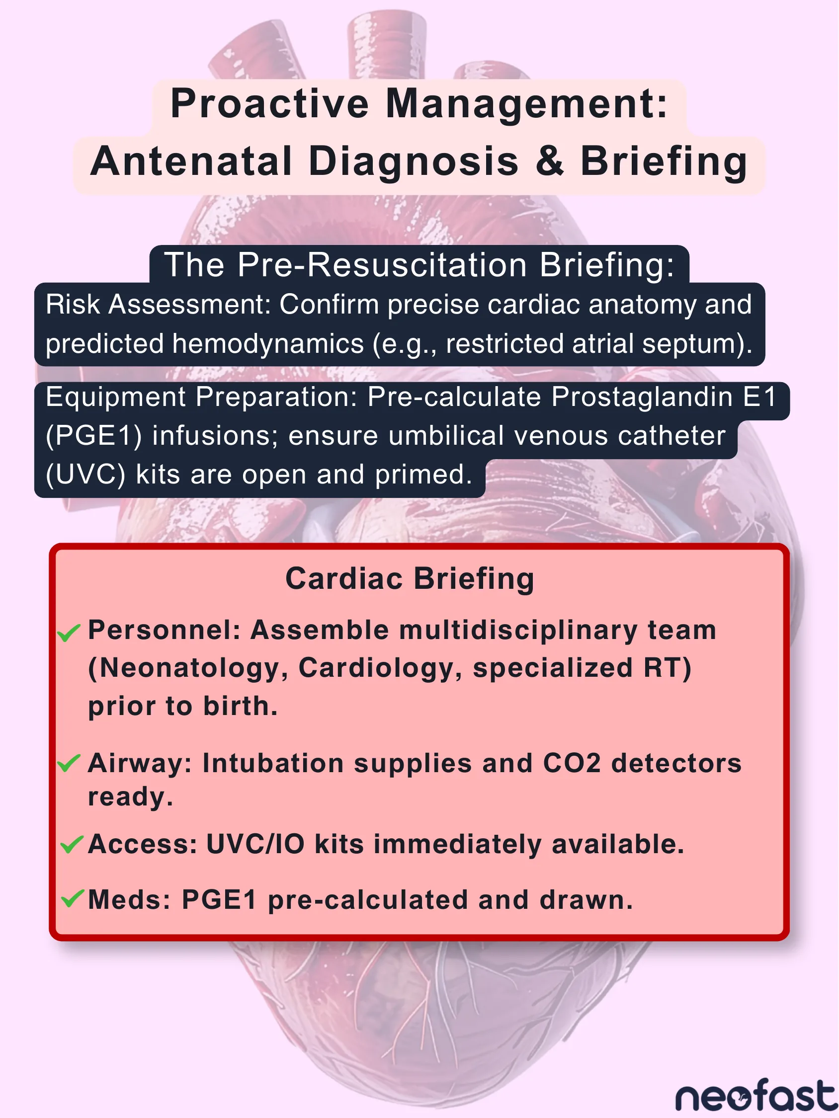
The Pre-Resuscitation Briefing:
- Risk Assessment: Confirm precise cardiac anatomy and predicted hemodynamics (e.g., restricted atrial septum).
- Equipment Preparation: Pre-calculate Prostaglandin E1 (PGE1) infusions; ensure umbilical venous catheter (UVC) kits are open and primed.
Cardiac Briefing
- Personnel: Assemble multidisciplinary team (Neonatology, Cardiology, specialized RT) prior to birth.
- Airway: Intubation supplies and CO2 detectors ready.
- Access: UVC/IO kits immediately available.
- Meds: PGE1 pre-calculated and drawn.
PGE1 Dosing Calculator
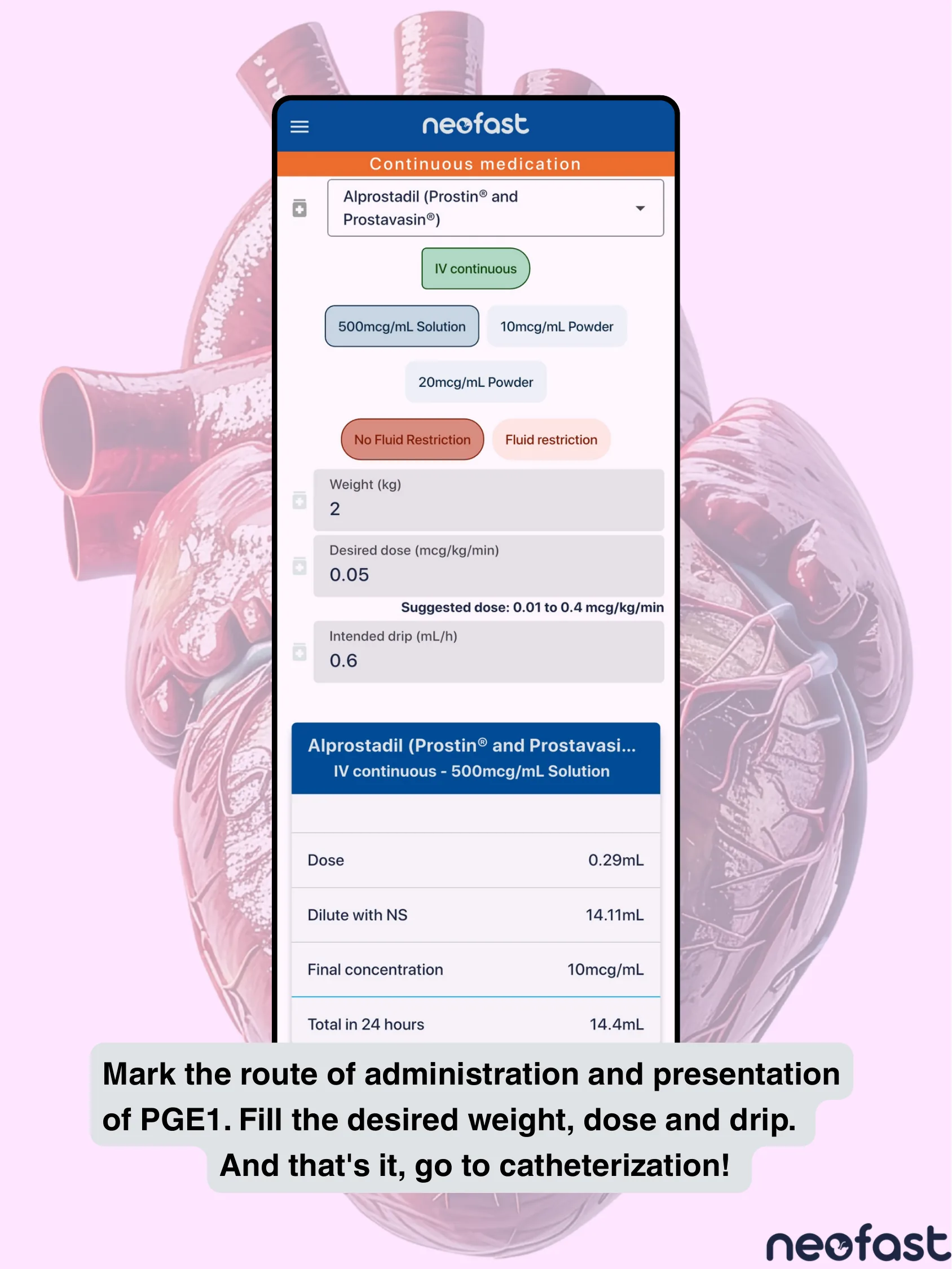
Continuous Medication: Alprostadil (Prostin® and Prostavasin®)
- Route: IV continuous
- Presentation options: 500mcg/mL Solution, 10mcg/mL Powder, 20mcg/mL Powder
- Fluid options: No Fluid Restriction, Fluid restriction
- Weight (kg): 2
- Desired dose (mcg/kg/min): 0.05
- Suggested dose: 0.01 to 0.4 mcg/kg/min
- Intended drip (mL/h): 0.6
Alprostadil (Prostin® and Prostavasin…) IV continuous – 500mcg/mL Solution
| Dose | 0.29mL |
| Dilute with NS | 14.11mL |
| Final concentration | 10mcg/mL |
| Total in 24 hours | 14.4mL |
Mark the route of administration and presentation of PGE1. Fill the desired weight, dose and drip. And that’s it, go to catheterization!
Ductal-Dependent Lesions
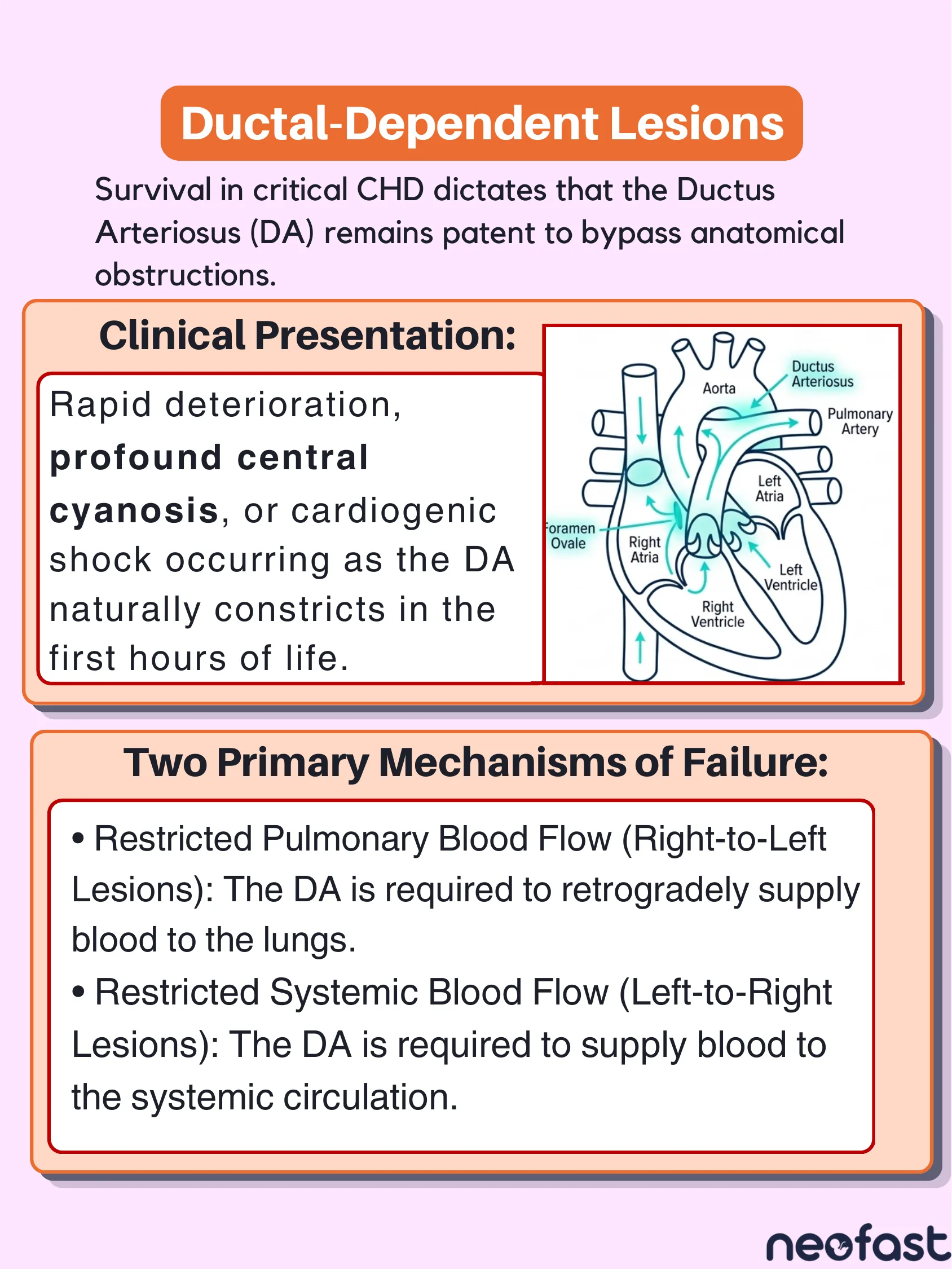
Survival in critical CHD dictates that the Ductus Arteriosus (DA) remains patent to bypass anatomical obstructions.
Clinical Presentation:
Rapid deterioration, profound central cyanosis, or cardiogenic shock occurring as the DA naturally constricts in the first hours of life.
Two Primary Mechanisms of Failure:
- Restricted Pulmonary Blood Flow (Right-to-Left Lesions): The DA is required to retrogradely supply blood to the lungs.
- Restricted Systemic Blood Flow (Left-to-Right Lesions): The DA is required to supply blood to the systemic circulation.
Differentiating Ductal-Dependent Pathologies
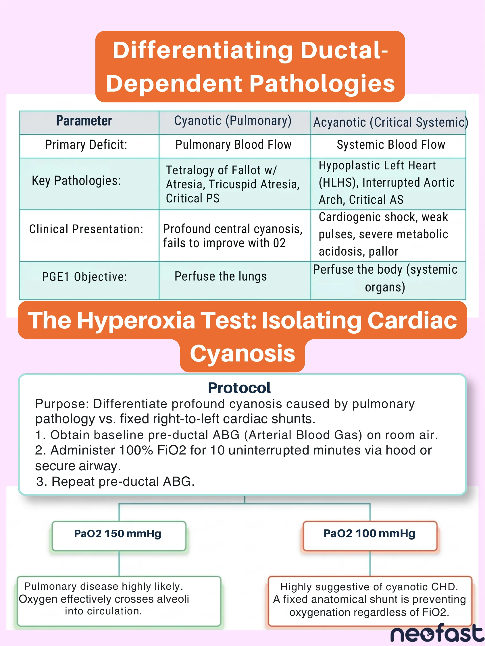
| Parameter | Cyanotic (Pulmonary) | Acyanotic (Critical Systemic) |
|---|---|---|
| Primary Deficit: | Pulmonary Blood Flow | Systemic Blood Flow |
| Key Pathologies: | Tetralogy of Fallot w/ Atresia, Tricuspid Atresia, Critical PS | Hypoplastic Left Heart (HLHS), Interrupted Aortic Arch, Critical AS |
| Clinical Presentation: | Profound central cyanosis, fails to improve with O2 | Cardiogenic shock, weak pulses, severe metabolic acidosis, pallor |
| PGE1 Objective: | Perfuse the lungs | Perfuse the body (systemic organs) |
The Hyperoxia Test: Isolating Cardiac Cyanosis
Protocol
Purpose: Differentiate profound cyanosis caused by pulmonary pathology vs. fixed right-to-left cardiac shunts.
- 1. Obtain baseline pre-ductal ABG (Arterial Blood Gas) on room air.
- 2. Administer 100% FiO2 for 10 uninterrupted minutes via hood or secure airway.
- 3. Repeat pre-ductal ABG.
- PaO2 150 mmHg: Pulmonary disease highly likely. Oxygen effectively crosses alveoli into circulation.
- PaO2 100 mmHg: Highly suggestive of cyanotic CHD. A fixed anatomical shunt is preventing oxygenation regardless of FiO2.
Adapting the NRP Algorithm for Suspected CHD
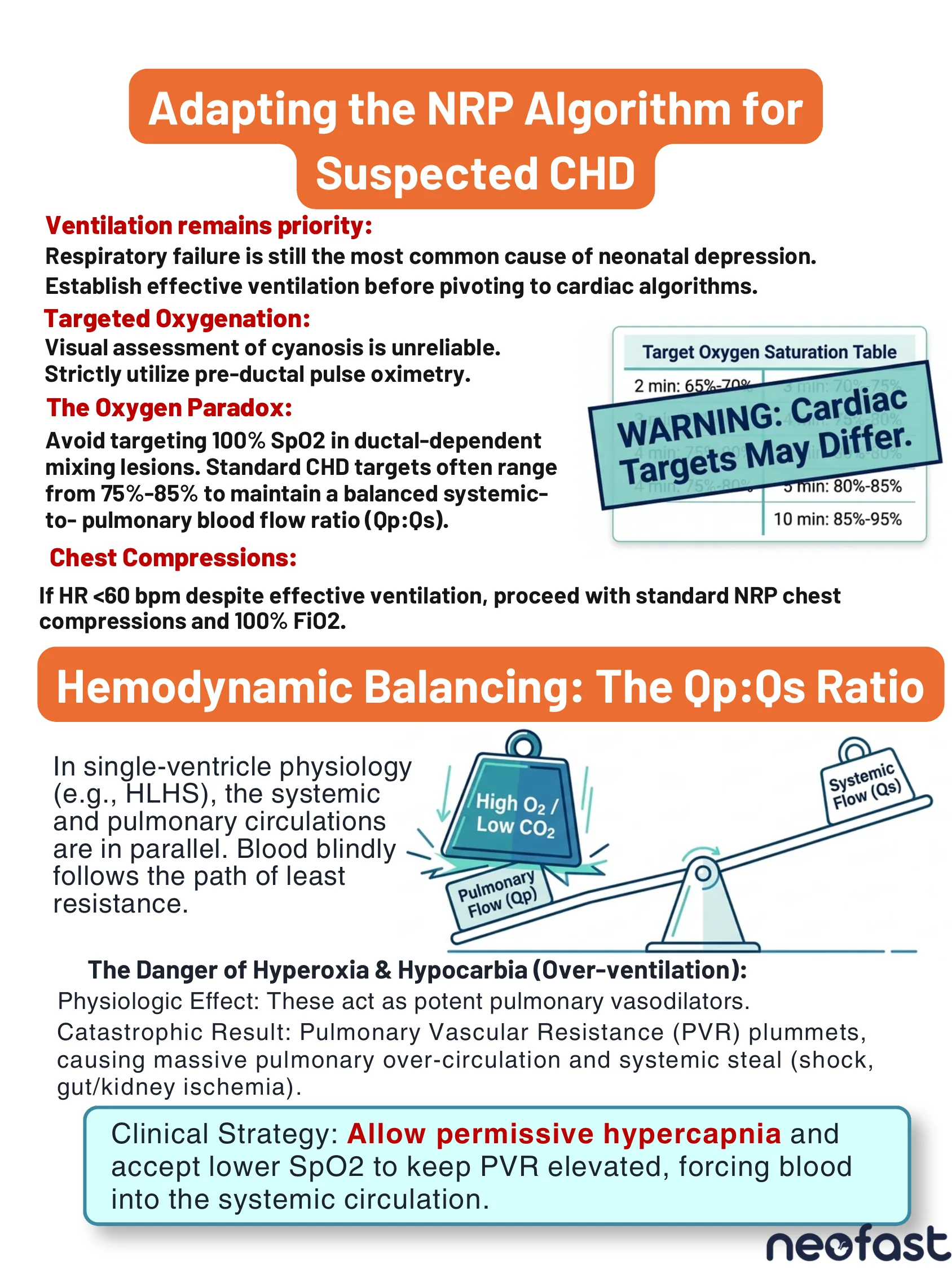
Ventilation remains priority:
Respiratory failure is still the most common cause of neonatal depression. Establish effective ventilation before pivoting to cardiac algorithms.
Targeted Oxygenation:
Visual assessment of cyanosis is unreliable. Strictly utilize pre-ductal pulse oximetry.
The Oxygen Paradox:
Avoid targeting 100% SpO2 in ductal-dependent mixing lesions. Standard CHD targets often range from 75%-85% to maintain a balanced systemic-to-pulmonary blood flow ratio (Qp:Qs).
WARNING: Cardiac Targets May Differ.
Target Oxygen Saturation Table
- 2 min: 65%-70%
- 3 min: 70%-75%
- 4 min: 75%-80%
- 5 min: 80%-85%
- 10 min: 85%-95%
Chest Compressions:
If HR <60 bpm despite effective ventilation, proceed with standard NRP chest compressions and 100% FiO2.
Hemodynamic Balancing: The Qp:Qs Ratio
In single-ventricle physiology (e.g., HLHS), the systemic and pulmonary circulations are in parallel. Blood blindly follows the path of least resistance.
The Danger of Hyperoxia & Hypocarbia (Over-ventilation):
Physiologic Effect: These act as potent pulmonary vasodilators.
Catastrophic Result: Pulmonary Vascular Resistance (PVR) plummets, causing massive pulmonary over-circulation and systemic steal (shock, gut/kidney ischemia).
Clinical Strategy: Allow permissive hypercapnia and accept lower SpO2 to keep PVR elevated, forcing blood into the systemic circulation.
Pharmacotherapy: Prostaglandin E1 (PGE1) Protocol
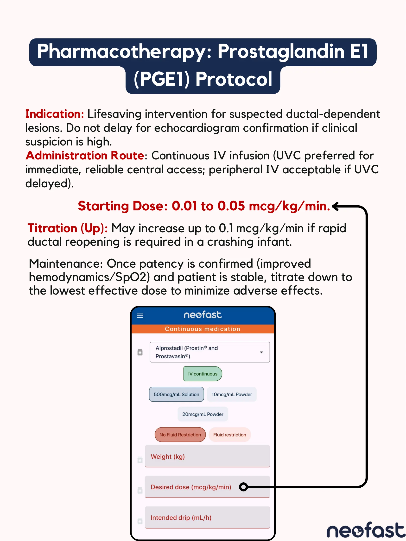
Indication: Lifesaving intervention for suspected ductal-dependent lesions. Do not delay for echocardiogram confirmation if clinical suspicion is high.
Administration Route: Continuous IV infusion (UVC preferred for immediate, reliable central access; peripheral IV acceptable if UVC delayed).
Starting Dose: 0.01 to 0.05 mcg/kg/min.
Titration (Up): May increase up to 0.1 mcg/kg/min if rapid ductal reopening is required in a crashing infant.
Maintenance: Once patency is confirmed (improved hemodynamics/SpO2) and patient is stable, titrate down to the lowest effective dose to minimize adverse effects.
Continuous medication: Alprostadil (Prostin® and Prostavasin®)
- IV continuous
- 500mcg/mL Solution / 10mcg/mL Powder / 20mcg/mL Powder
- No Fluid Restriction / Fluid restriction
- Weight (kg)
- Desired dose (mcg/kg/min)
- Intended drip (mL/h)
Anticipating PGE1 Complications
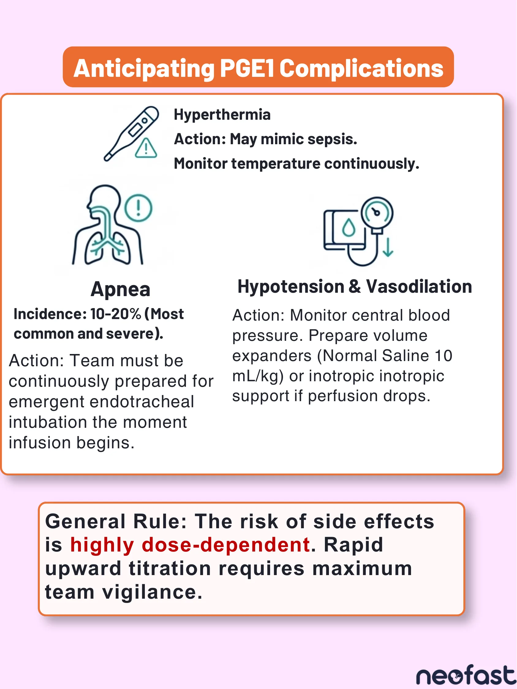
Hyperthermia
Action: May mimic sepsis. Monitor temperature continuously.
Apnea
Incidence: 10-20% (Most common and severe).
Action: Team must be continuously prepared for emergent endotracheal intubation the moment infusion begins.
Hypotension & Vasodilation
Action: Monitor central blood pressure. Prepare volume expanders (Normal Saline 10 mL/kg) or inotropic support if perfusion drops.
General Rule: The risk of side effects is highly dose-dependent. Rapid upward titration requires maximum team vigilance.
Key Takeaways
Key Takeaways
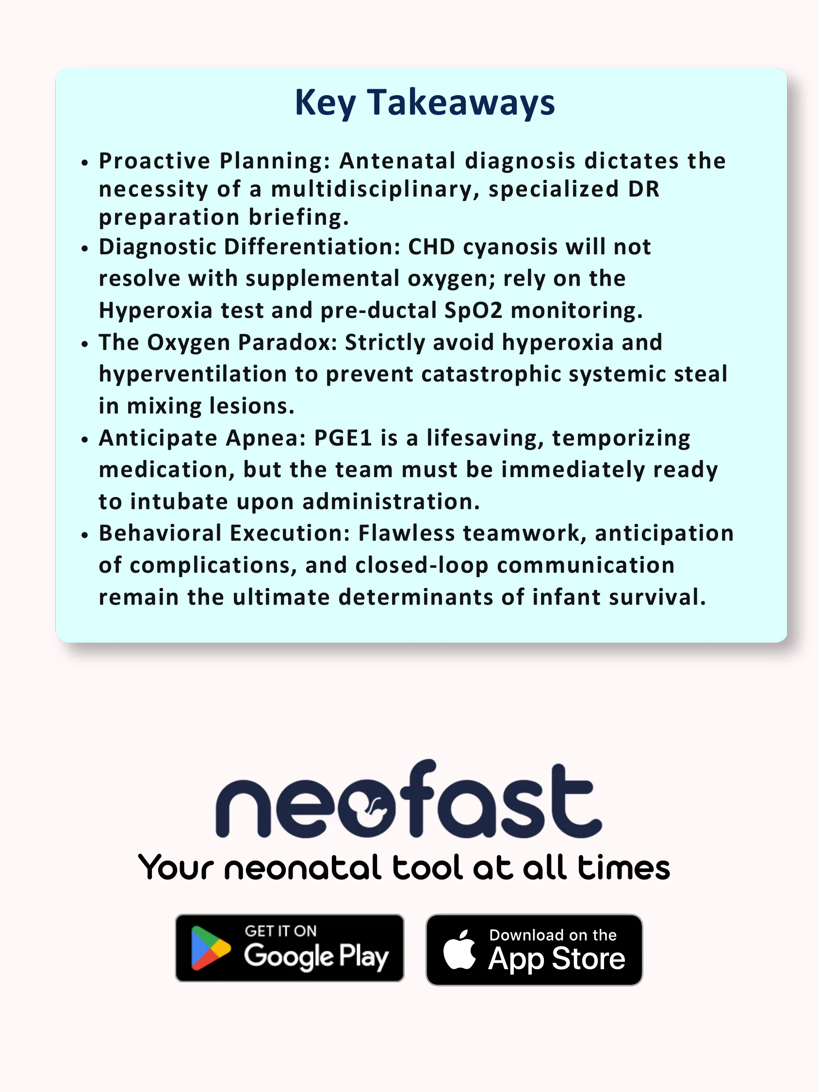
- Proactive Planning: Antenatal diagnosis dictates the necessity of a multidisciplinary, specialized DR preparation briefing.
- Diagnostic Differentiation: CHD cyanosis will not resolve with supplemental oxygen; rely on the Hyperoxia test and pre-ductal SpO2 monitoring.
- The Oxygen Paradox: Strictly avoid hyperoxia and hyperventilation to prevent catastrophic systemic steal in mixing lesions.
- Anticipate Apnea: PGE1 is a lifesaving, temporizing medication, but the team must be immediately ready to intubate upon administration.
- Behavioral Execution: Flawless teamwork, anticipation of complications, and closed-loop communication remain the ultimate determinants of infant survival.
neofast
Your neonatal tool at all times
Frequently asked questions
What is the starting dose of PGE1 (Alprostadil) for suspected ductal-dependent CHD?
Starting dose is 0.01 to 0.05 mcg/kg/min IV continuous infusion, titratable up to 0.1 mcg/kg/min if rapid ductal reopening is needed in a crashing infant, then titrated down to the lowest effective dose once stable.
What is the most common complication of PGE1 infusion in neonates?
Apnea occurs in 10-20% of infants, making it the most common and most severe complication; the team must be continuously prepared for emergent endotracheal intubation the moment the infusion begins. Other complications include hyperthermia (which may mimic sepsis) and hypotension/vasodilation, managed with central BP monitoring, normal saline 10 mL/kg volume expansion, or inotropic support.
How does the Hyperoxia Test help differentiate cyanotic CHD from pulmonary disease?
After obtaining a baseline pre-ductal ABG on room air and administering 100% FiO2 for 10 uninterrupted minutes, a repeat PaO2 of 150 mmHg suggests pulmonary disease (oxygen crosses alveoli effectively), whereas a PaO2 of 100 mmHg is highly suggestive of cyanotic CHD due to a fixed anatomical shunt preventing oxygenation regardless of FiO2.
What SpO2 targets should be used during resuscitation of infants with suspected ductal-dependent mixing lesions?
Avoid targeting 100% SpO2; use pre-ductal pulse oximetry with targets of 65%-70% at 2 min, 70%-75% at 3 min, 75%-80% at 4 min, 80%-85% at 5 min, and 85%-95% at 10 min, since standard CHD targets of 75%-85% maintain a balanced Qp:Qs ratio.
Why should hyperoxia and hyperventilation be avoided in single-ventricle physiology like HLHS?
Hyperoxia and hypocarbia act as potent pulmonary vasodilators, causing PVR to plummet, which leads to massive pulmonary over-circulation and systemic steal (shock, gut/kidney ischemia); the strategy instead is to allow permissive hypercapnia and accept lower SpO2 to keep PVR elevated and force blood into the systemic circulation.