Key takeaways
- In Japanese NICUs, survival at 22 weeks gestation reaches approximately 60% (NRNJ Registry Data), rising to 78% at 23 weeks, 84% at 24 weeks, 89% at 25 weeks, 94% at 26 weeks, and 97% at 27 weeks.
- High-Frequency Oscillatory Ventilation (HFOV) is deployed as a proactive, lung-protective first-line strategy rather than a rescue therapy, and is considered a primary driver of 22-week viability.
- Initial HFOV settings should be MAP set 4-5 cmH2O above the CMV baseline (or 1.5x the CMV baseline, e.g., CMV MAP of 8 to HFOV MAP of 12), Amplitude (Delta P) default of 20 cmH2O, and Frequency centered at 12 Hz.
- The 22-23 week trachea requires a 2.0mm ETT, which causes severe dampening of pressure amplitudes and carries massive risk of fatal secretion blockages, requiring continuous monitoring of graphic waves and pressure amplitude trends.
- For RDS, target VThf is 1.0-2.0 mL/kg with Hz >12; for severe Chronic Lung Disease (CLD), target VThf is 2.0-3.0 mL/kg with Hz <12, and Delta P should be adjusted to maintain pCO2 at 50-60 mmHg.
References
- KUSUDA, Satoshi; NAKANISHI, Hidehiko; ISAYAMA, Tetsuya (Eds.). Neonatal Intensive Care for Extremely Preterm Infants: Japanese NICU Practices to Prevent Mortality and Morbidities. Elsevier/Academic Press, 2024
Japanese First-Line High-Frequency Oscillatory Ventilation Protocols for the Extreme Preterm Infants
The 22-Week Clinical Playbook
Source: KUSUDA, Satoshi; NAKANISHI, Hidehiko; ISAYAMA, Tetsuya (Eds.). Neonatal Intensive Care for Extremely Preterm Infants: Japanese NICU Practices to Prevent Mortality and Morbidities. Elsevier/Academic Press, 2024
Redefining survival at the edge of viability
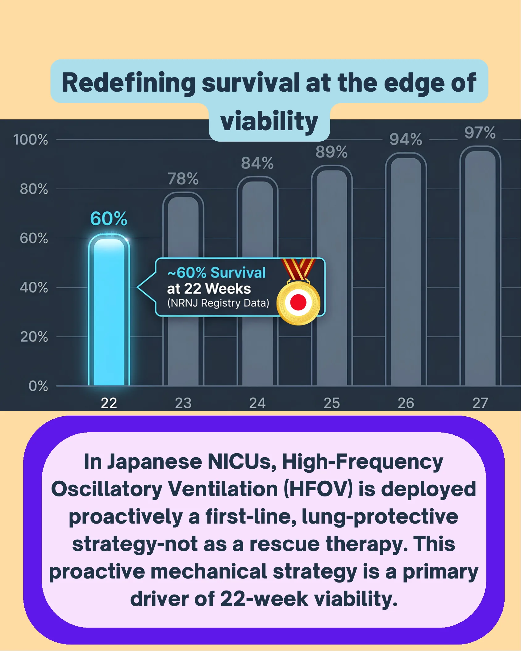
Redefining survival at the edge of viability
| Gestational Week | Survival Rate |
|---|---|
| 22 | 60% |
| 23 | 78% |
| 24 | 84% |
| 25 | 89% |
| 26 | 94% |
| 27 | 97% |
~60% Survival at 22 Weeks (NRNJ Registry Data)
In Japanese NICUs, High-Frequency Oscillatory Ventilation (HFOV) is deployed proactively a first-line, lung-protective strategy-not as a rescue therapy. This proactive mechanical strategy is a primary driver of 22-week viability.
The physical limits of the canalicular lung
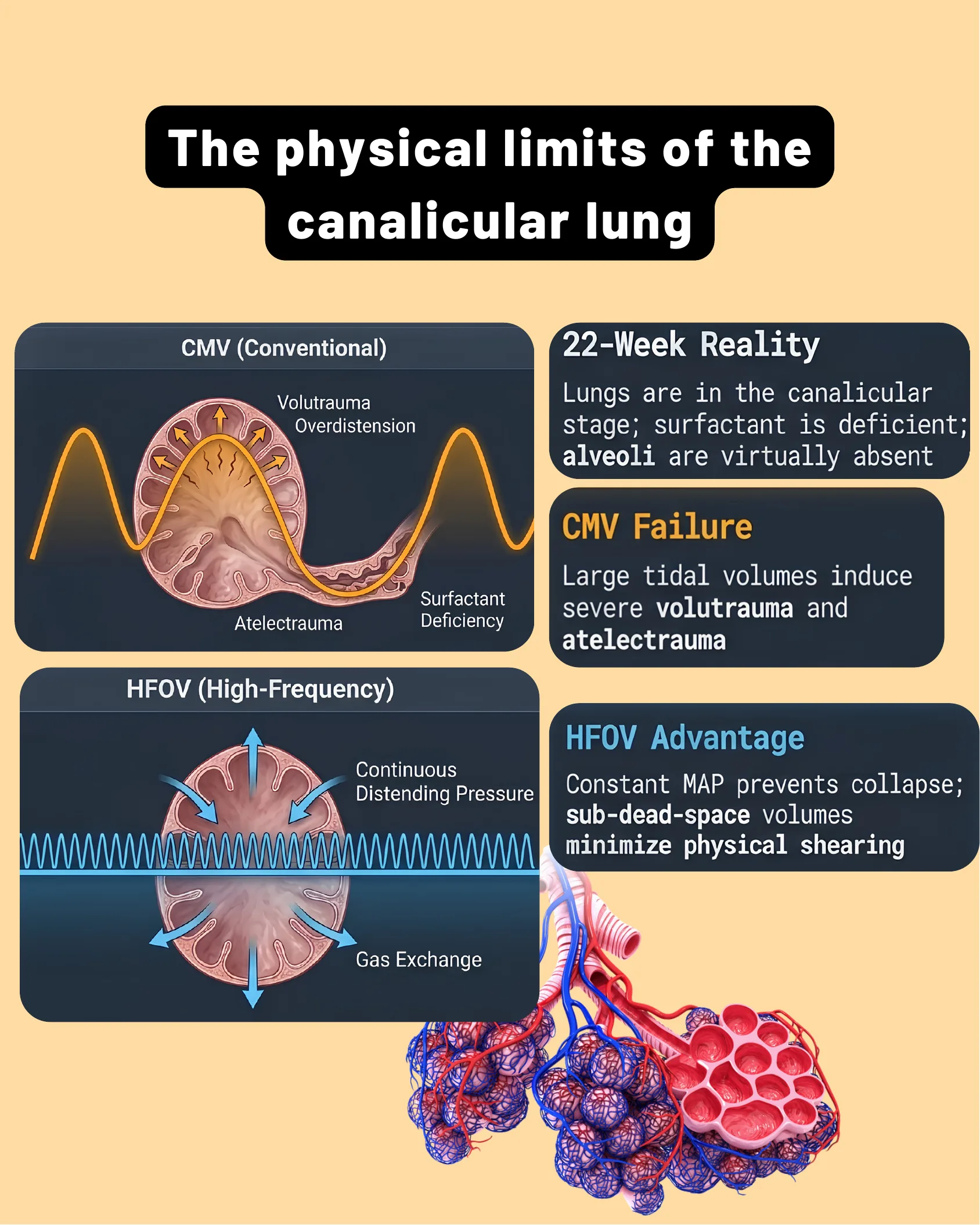
The physical limits of the canalicular lung
CMV (Conventional)
- Volutrauma / Overdistension
- Atelectrauma
- Surfactant Deficiency
HFOV (High-Frequency)
- Continuous Distending Pressure
- Gas Exchange
22-Week Reality
Lungs are in the canalicular stage; surfactant is deficient; alveoli are virtually absent
CMV Failure
Large tidal volumes induce severe volutrauma and atelectrauma
HFOV Advantage
Constant MAP prevents collapse; sub-dead-space volumes minimize physical shearing
The Core HFOV Interface
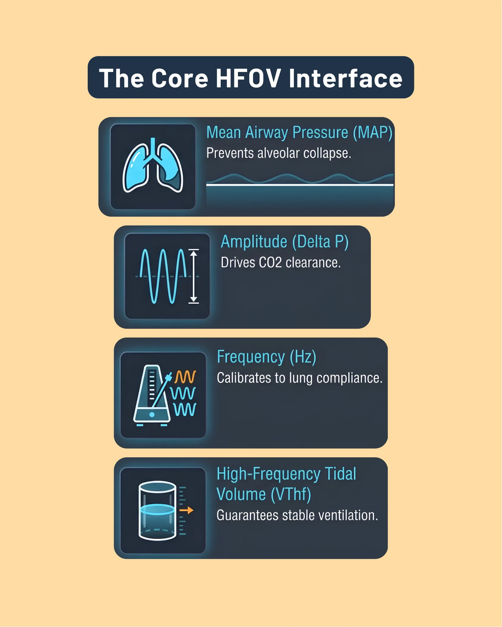
The Core HFOV Interface
- Mean Airway Pressure (MAP) – Prevents alveolar collapse.
- Amplitude (Delta P) – Drives CO2 clearance.
- Frequency (Hz) – Calibrates to lung compliance.
- High-Frequency Tidal Volume (VThf) – Guarantees stable ventilation.
Establishing baseline volume
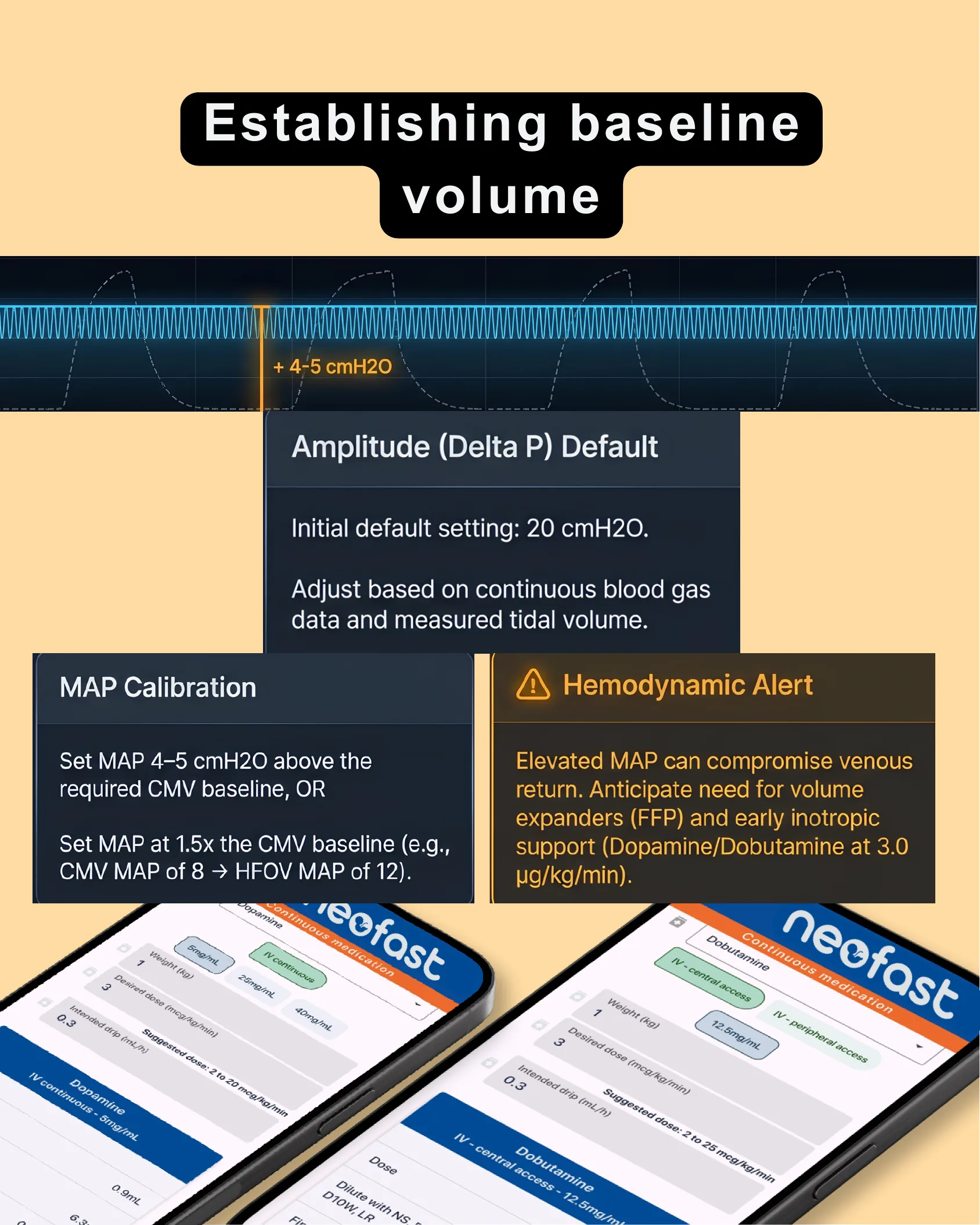
Establishing baseline volume
+ 4-5 cmH2O
Amplitude (Delta P) Default
Initial default setting: 20 cmH2O.
Adjust based on continuous blood gas data and measured tidal volume.
MAP Calibration
- Set MAP 4-5 cmH2O above the required CMV baseline, OR
- Set MAP at 1.5x the CMV baseline (e.g., CMV MAP of 8 → HFOV MAP of 12).
Hemodynamic Alert
Elevated MAP can compromise venous return. Anticipate need for volume expanders (FFP) and early inotropic support (Dopamine/Dobutamine at 3.0 µg/kg/min).
Calibrating for compliance
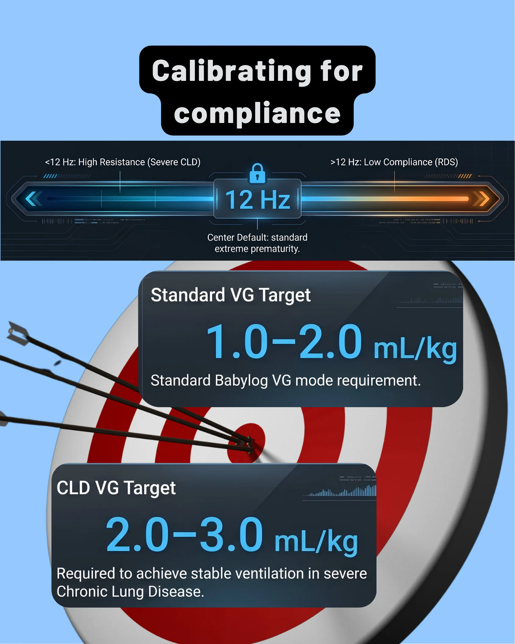
Calibrating for compliance
12 Hz – Center Default: standard extreme prematurity.
- <12 Hz: High Resistance (Severe CLD)
- >12 Hz: Low Compliance (RDS)
Standard VG Target
1.0-2.0 mL/kg – Standard Babylog VG mode requirement.
CLD VG Target
2.0-3.0 mL/kg – Required to achieve stable ventilation in severe Chronic Lung Disease.
The anatomical bottleneck: ETT Physics
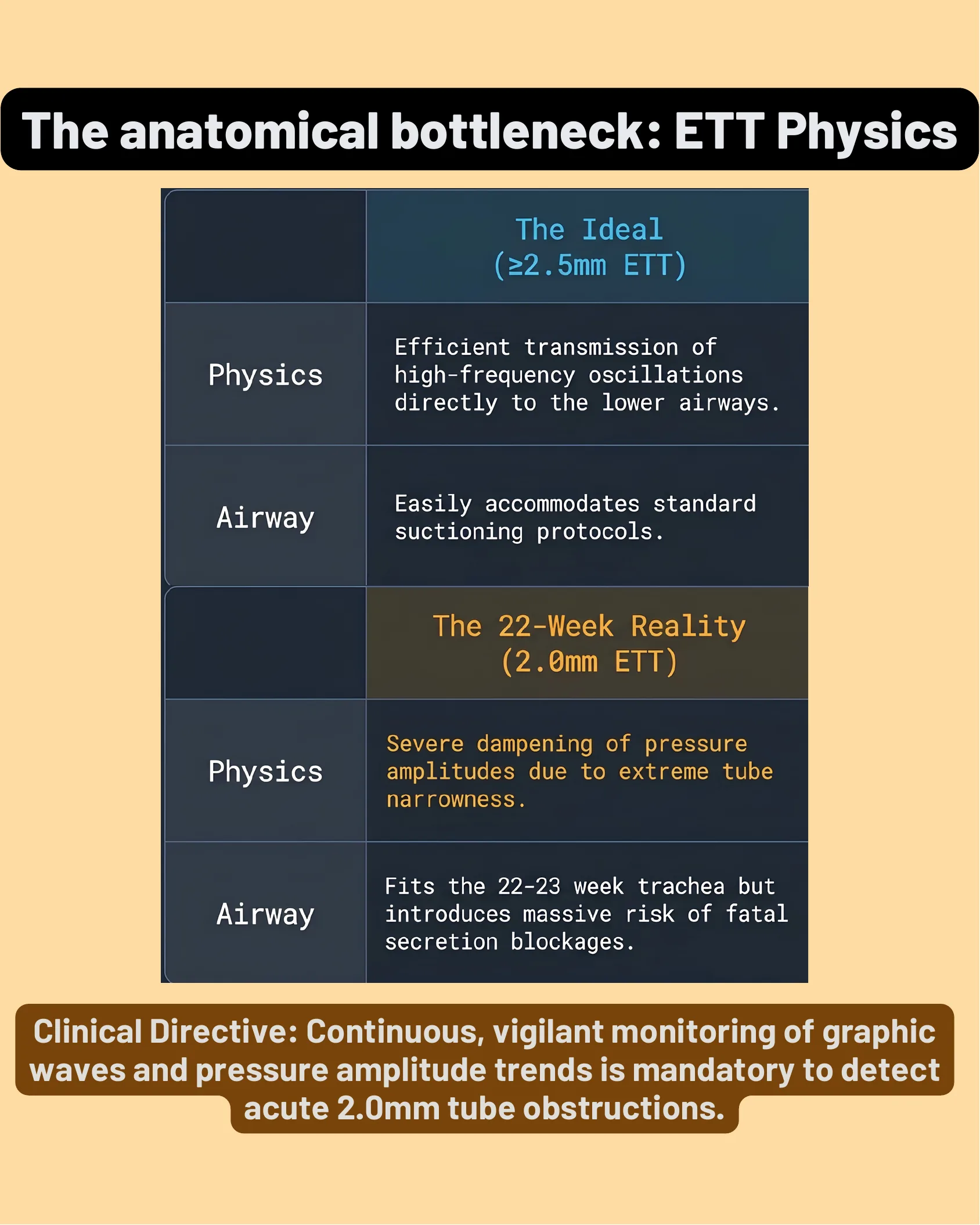
The anatomical bottleneck: ETT Physics
| The Ideal (≥2.5mm ETT) | |
|---|---|
| Physics | Efficient transmission of high-frequency oscillations directly to the lower airways. |
| Airway | Easily accommodates standard suctioning protocols. |
| The 22-Week Reality (2.0mm ETT) | |
|---|---|
| Physics | Severe dampening of pressure amplitudes due to extreme tube narrowness. |
| Airway | Fits the 22-23 week trachea but introduces massive risk of fatal secretion blockages. |
Clinical Directive: Continuous, vigilant monitoring of graphic waves and pressure amplitude trends is mandatory to detect acute 2.0mm tube obstructions.
The 22-Week HFOV Calibration Algorithm
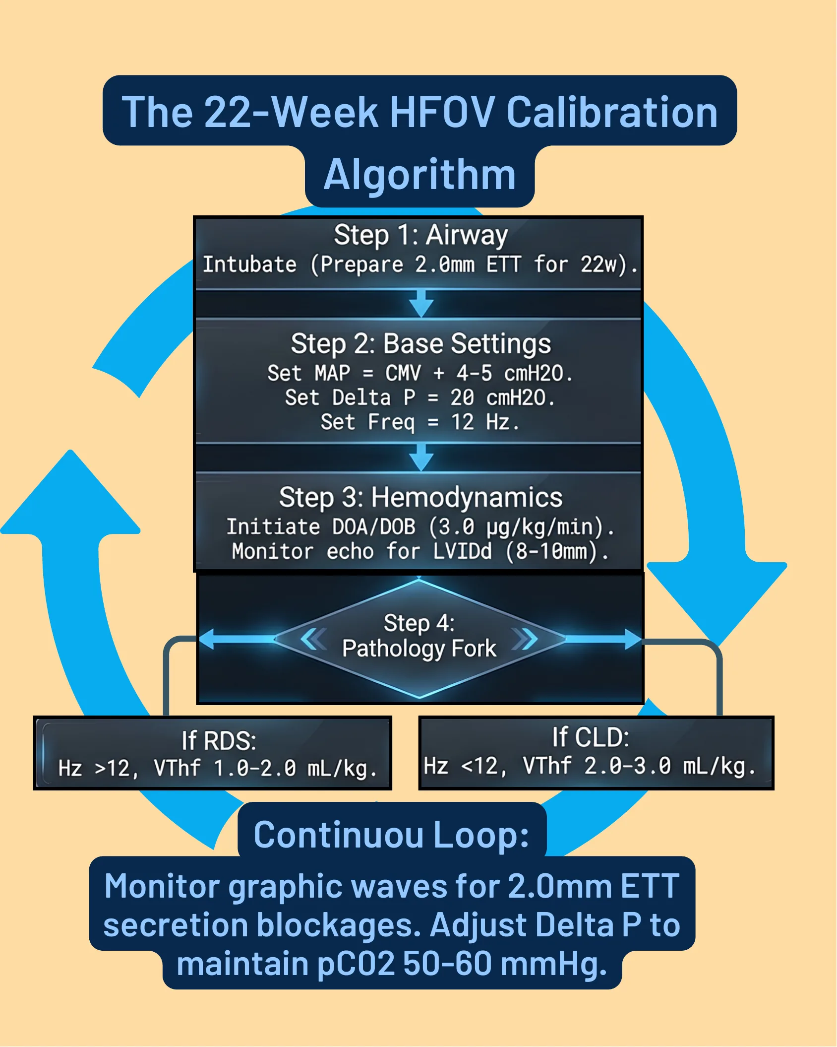
The 22-Week HFOV Calibration Algorithm
- Step 1: Airway – Intubate (Prepare 2.0mm ETT for 22w).
- Step 2: Base Settings – Set MAP = CMV + 4-5 cmH2O. Set Delta P = 20 cmH2O. Set Freq = 12 Hz.
- Step 3: Hemodynamics – Initiate DOA/DOB (3.0 µg/kg/min). Monitor echo for LVIDd (8-10mm).
- Step 4: Pathology Fork
- If RDS: Hz >12, VThf 1.0-2.0 mL/kg.
- If CLD: Hz <12, VThf 2.0-3.0 mL/kg.
Continuous Loop: Monitor graphic waves for 2.0mm ETT secretion blockages. Adjust Delta P to maintain pCO2 50-60 mmHg.
Neofast App Main Menu

Neofast
- Venous hydration
- Continuous medication
- Medicines
- Intubation
- Other calculations and scores
- Procedures
Mechanical ventilation coming soon!
Download on the App Store
Get it on Google Play
Frequently asked questions
What is the survival rate for extremely preterm infants born at 22 weeks gestation in Japanese NICUs?
According to NRNJ Registry Data, survival at 22 weeks gestation is approximately 60%, increasing to 78% at 23 weeks, 84% at 24 weeks, 89% at 25 weeks, 94% at 26 weeks, and 97% at 27 weeks.
How should MAP be set when initiating HFOV from CMV in extremely preterm infants?
Set MAP either 4-5 cmH2O above the required CMV baseline, or at 1.5x the CMV baseline (for example, a CMV MAP of 8 becomes an HFOV MAP of 12).
What is the default amplitude (Delta P) and frequency setting for HFOV in the 22-week protocol?
The initial default Amplitude (Delta P) setting is 20 cmH2O, adjusted based on continuous blood gas data and measured tidal volume. The center default Frequency is 12 Hz, which is calibrated to lung compliance: less than 12 Hz for high resistance (severe CLD) and greater than 12 Hz for low compliance (RDS).
Why is a 2.0mm ETT risky in 22-week infants on HFOV?
A 2.0mm ETT fits the 22-23 week trachea but causes severe dampening of pressure amplitudes due to extreme tube narrowness and introduces massive risk of fatal secretion blockages, requiring mandatory continuous monitoring of graphic waves and pressure amplitude trends to detect acute obstructions.
What hemodynamic support should be anticipated when elevating MAP on HFOV?
Elevated MAP can compromise venous return, so clinicians should anticipate the need for volume expanders (FFP) and early inotropic support with Dopamine/Dobutamine at 3.0 µg/kg/min, while monitoring echo for LVIDd (8-10mm).