References
- Guia de Bolso de Neonatologia (3ª Edição, Santa Casa SP)
- Klaus & Fanaroff’s Care of the High-Risk Neonate (8th Ed)
- Guidelines for Acute Care of the Neonate (2025-26, Baylor College of Medicine)
- SOPERJ Neonatology Guidelines (2022)
Key takeaways
- Hypophosphatemia affects up to 60% of premature infants on parenteral nutrition and is a critical bottleneck for ATP synthesis and bone mineralization.
- The normal neonatal phosphate range is 4.5–9.5 mg/dL; moderate hypophosphatemia is 1.5–2.5 mg/dL and severe is below 1.5 mg/dL.
- 80% of fetal calcium and phosphorus accretion occurs in the third trimester, so preterm infants are born with profound mineral deficits.
Neonatal Hypophosphatemia: The Clinical Blueprint
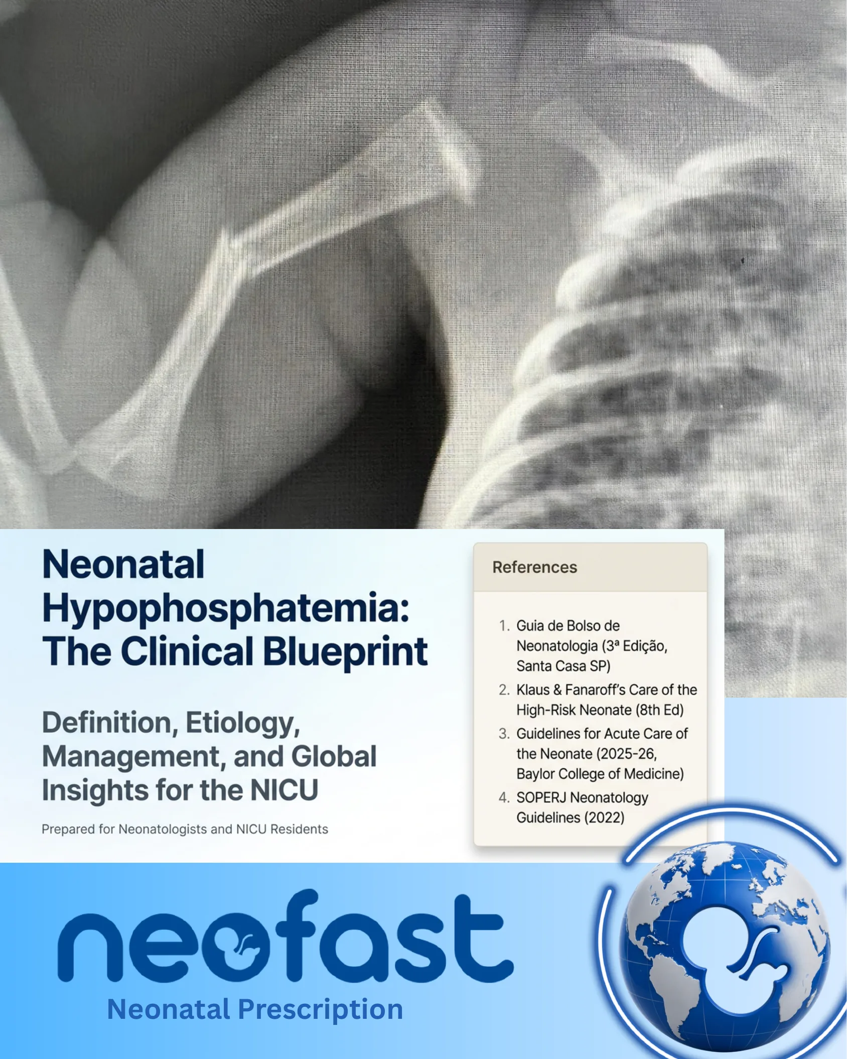
Neonatal Hypophosphatemia: The Clinical Blueprint
Definition, Etiology, Management, and Global Insights for the NICU
Prepared for Neonatologists and NICU Residents
References
- Guia de Bolso de Neonatologia (3ª Edição, Santa Casa SP)
- Klaus & Fanaroff’s Care of the High-Risk Neonate (8th Ed)
- Guidelines for Acute Care of the Neonate (2025-26, Baylor College of Medicine)
- SOPERJ Neonatology Guidelines (2022)
neofast — Neonatal Prescription
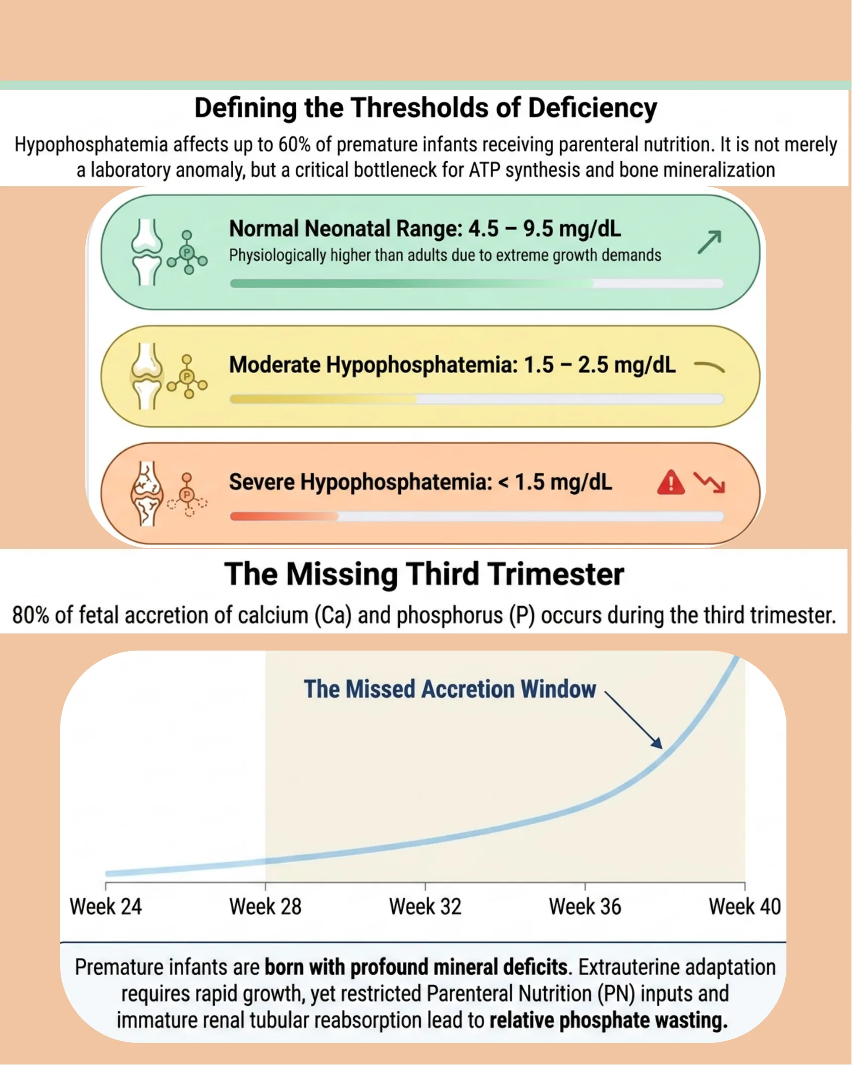
Defining the Thresholds of Deficiency
Defining the Thresholds of Deficiency
Hypophosphatemia affects up to 60% of premature infants receiving parenteral nutrition. It is not merely a laboratory anomaly, but a critical bottleneck for ATP synthesis and bone mineralization.
- Normal Neonatal Range: 4.5 – 9.5 mg/dL — Physiologically higher than adults due to extreme growth demands
- Moderate Hypophosphatemia: 1.5 – 2.5 mg/dL
- Severe Hypophosphatemia: < 1.5 mg/dL
The Missing Third Trimester
80% of fetal accretion of calcium (Ca) and phosphorus (P) occurs during the third trimester.
The Missed Accretion Window — chart spanning Week 24, Week 28, Week 32, Week 36, Week 40, showing an exponential increase in mineral accretion approaching term.
Premature infants are born with profound mineral deficits. Extrauterine adaptation requires rapid growth, yet restricted Parenteral Nutrition (PN) inputs and immature renal tubular reabsorption lead to relative phosphate wasting.
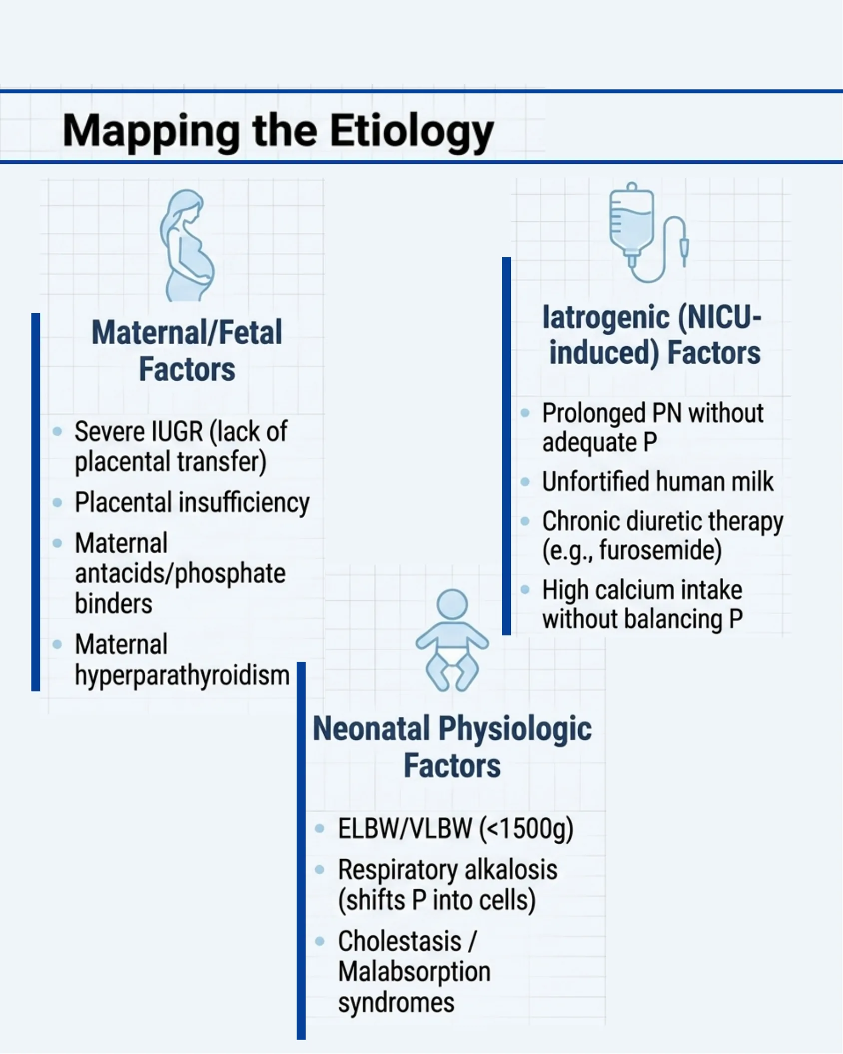
Mapping the Etiology
Mapping the Etiology
Maternal/Fetal Factors
- Severe IUGR (lack of placental transfer)
- Placental insufficiency
- Maternal antacids/phosphate binders
- Maternal hyperparathyroidism
Neonatal Physiologic Factors
- ELBW/VLBW (<1500g)
- Respiratory alkalosis (shifts P into cells)
- Cholestasis / Malabsorption syndromes
Iatrogenic (NICU-induced) Factors
- Prolonged PN without adequate P
- Unfortified human milk
- Chronic diuretic therapy (e.g., furosemide)
- High calcium intake without balancing P
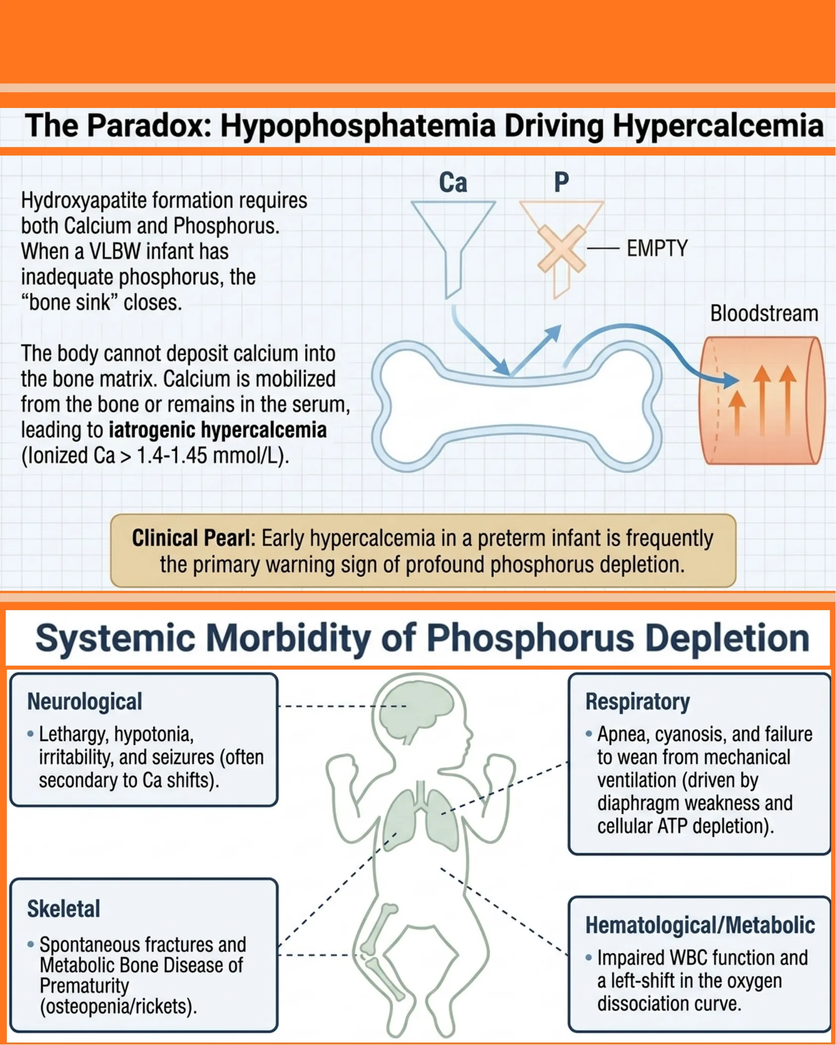
The Paradox: Hypophosphatemia Driving Hypercalcemia
The Paradox: Hypophosphatemia Driving Hypercalcemia
Hydroxyapatite formation requires both Calcium and Phosphorus. When a VLBW infant has inadequate phosphorus, the “bone sink” closes.
The body cannot deposit calcium into the bone matrix. Calcium is mobilized from the bone or remains in the serum, leading to iatrogenic hypercalcemia (Ionized Ca > 1.4-1.45 mmol/L).
Clinical Pearl: Early hypercalcemia in a preterm infant is frequently the primary warning sign of profound phosphorus depletion.
Systemic Morbidity of Phosphorus Depletion
- Neurological: Lethargy, hypotonia, irritability, and seizures (often secondary to Ca shifts).
- Respiratory: Apnea, cyanosis, and failure to wean from mechanical ventilation (driven by diaphragm weakness and cellular ATP depletion).
- Skeletal: Spontaneous fractures and Metabolic Bone Disease of Prematurity (osteopenia/rickets).
- Hematological/Metabolic: Impaired WBC function and a left-shift in the oxygen dissociation curve.
Comprehensive Diagnostic Surveillance
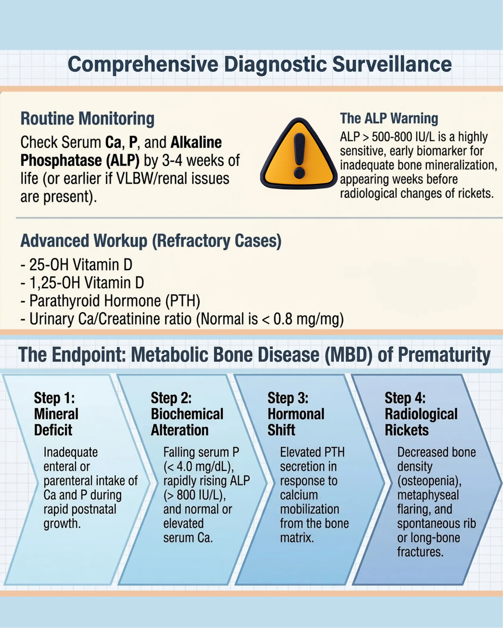
Comprehensive Diagnostic Surveillance
Routine Monitoring
Check Serum Ca, P, and Alkaline Phosphatase (ALP) by 3-4 weeks of life (or earlier if VLBW/renal issues are present).
The ALP Warning
ALP > 500-800 IU/L is a highly sensitive, early biomarker for inadequate bone mineralization, appearing weeks before radiological changes of rickets.
Advanced Workup (Refractory Cases)
- 25-OH Vitamin D
- 1,25-OH Vitamin D
- Parathyroid Hormone (PTH)
- Urinary Ca/Creatinine ratio (Normal is < 0.8 mg/mg)
The Endpoint: Metabolic Bone Disease (MBD) of Prematurity
- Step 1: Mineral Deficit — Inadequate enteral or parenteral intake of Ca and P during rapid postnatal growth.
- Step 2: Biochemical Alteration — Falling serum P (< 4.0 mg/dL), rapidly rising ALP (> 800 IU/L), and normal or elevated serum Ca.
- Step 3: Hormonal Shift — Elevated PTH secretion in response to calcium mobilization from the bone matrix.
- Step 4: Radiological Rickets — Decreased bone density (osteopenia), metaphyseal flaring, and spontaneous rib or long-bone fractures.
Parenteral Management: The Golden Ratios
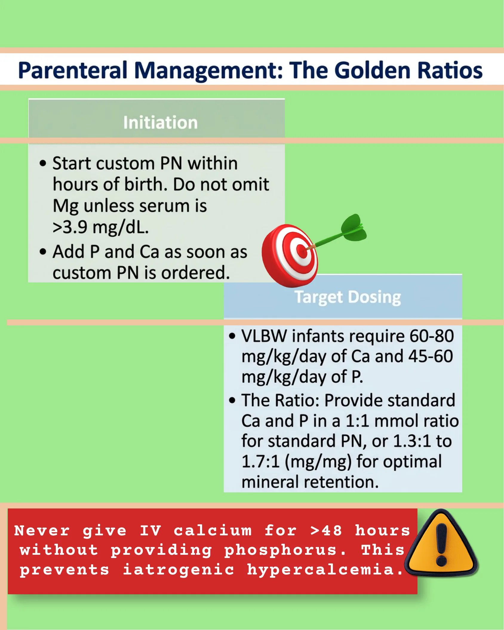
Parenteral Management: The Golden Ratios
Initiation
- Start custom PN within hours of birth. Do not omit Mg unless serum is >3.9 mg/dL.
- Add P and Ca as soon as custom PN is ordered.
Target Dosing
- VLBW infants require 60-80 mg/kg/day of Ca and 45-60 mg/kg/day of P.
- The Ratio: Provide standard Ca and P in a 1:1 mmol ratio for standard PN, or 1.3:1 to 1.7:1 (mg/mg) for optimal mineral retention.
Never give IV calcium for >48 hours without providing phosphorus. This prevents iatrogenic hypercalcemia.
Enteral Management & Fortification
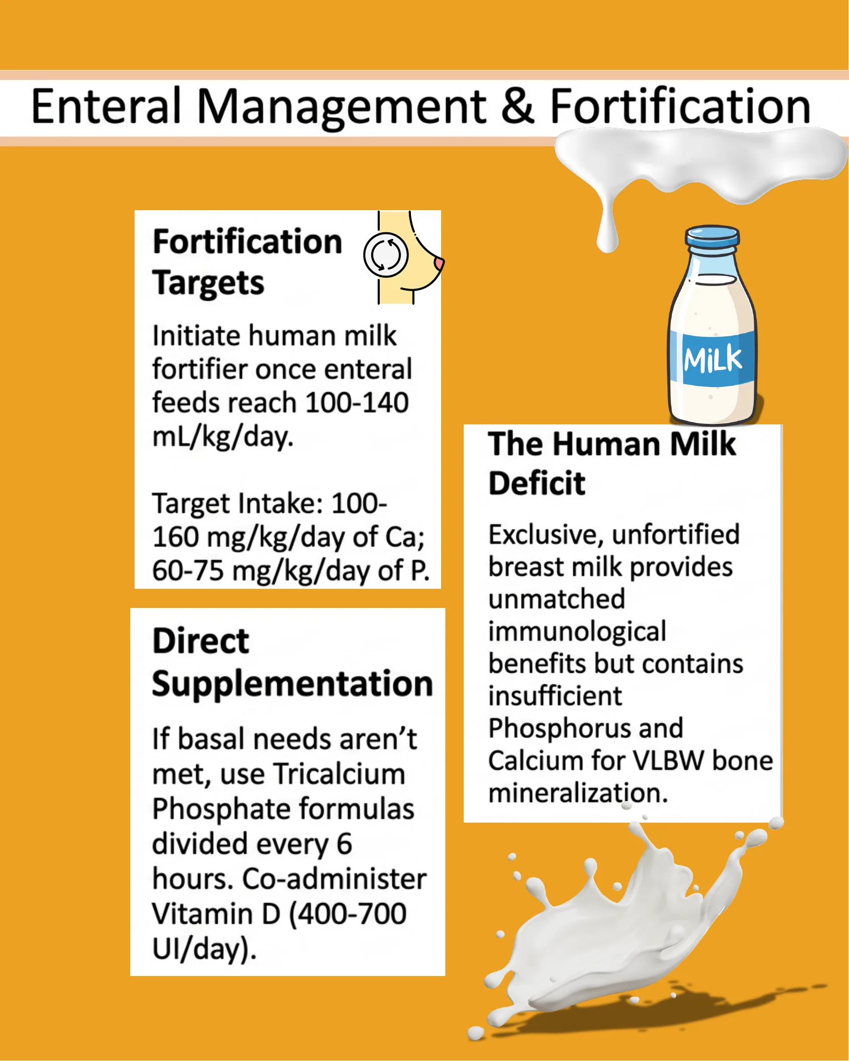
Enteral Management & Fortification
Fortification Targets
Initiate human milk fortifier once enteral feeds reach 100-140 mL/kg/day.
Target Intake: 100-160 mg/kg/day of Ca; 60-75 mg/kg/day of P.
The Human Milk Deficit
Exclusive, unfortified breast milk provides unmatched immunological benefits but contains insufficient Phosphorus and Calcium for VLBW bone mineralization.
Direct Supplementation
If basal needs aren’t met, use Tricalcium Phosphate formulas divided every 6 hours. Co-administer Vitamin D (400-700 UI/day).
Acute Correction Algorithm
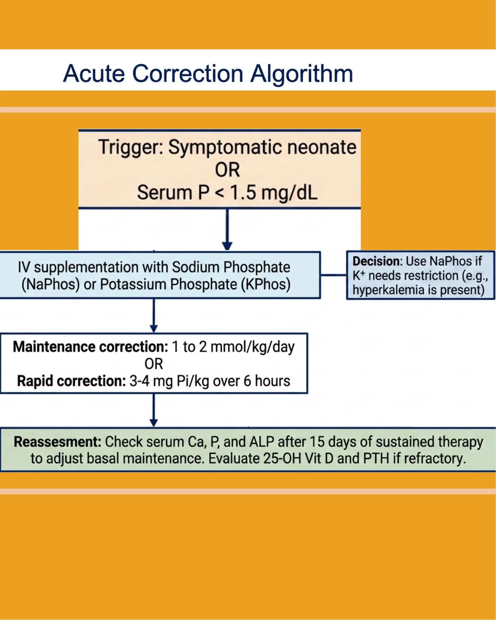
Acute Correction Algorithm
- Trigger: Symptomatic neonate OR Serum P < 1.5 mg/dL
- Action: IV supplementation with Sodium Phosphate (NaPhos) or Potassium Phosphate (KPhos)
- Decision: Use NaPhos if K+ needs restriction (e.g., hyperkalemia is present)
- Dosing:
- Maintenance correction: 1 to 2 mmol/kg/day
- OR Rapid correction: 3-4 mg Pi/kg over 6 hours
- Reassessment: Check serum Ca, P, and ALP after 15 days of sustained therapy to adjust basal maintenance. Evaluate 25-OH Vit D and PTH if refractory.
At the Frontier: Global Research & Emerging Insights
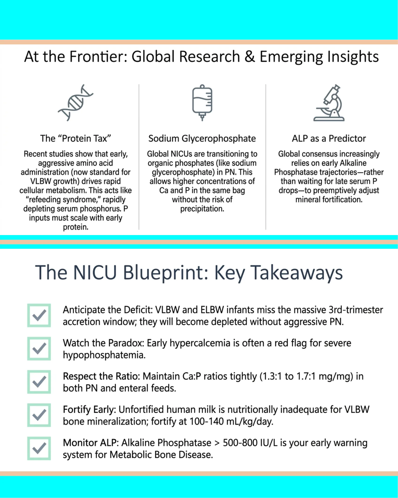
At the Frontier: Global Research & Emerging Insights
The “Protein Tax”
Recent studies show that early, aggressive amino acid administration (now standard for VLBW growth) drives rapid cellular metabolism. This acts like “refeeding syndrome,” rapidly depleting serum phosphorus. P inputs must scale with early protein.
Sodium Glycerophosphate
Global NICUs are transitioning to organic phosphates (like sodium glycerophosphate) in PN. This allows higher concentrations of Ca and P in the same bag without the risk of precipitation.
ALP as a Predictor
Global consensus increasingly relies on early Alkaline Phosphatase trajectories—rather than waiting for late serum P drops—to preemptively adjust mineral fortification.
The NICU Blueprint: Key Takeaways
- Anticipate the Deficit: VLBW and ELBW infants miss the massive 3rd-trimester accretion window; they will become depleted without aggressive PN.
- Watch the Paradox: Early hypercalcemia is often a red flag for severe hypophosphatemia.
- Respect the Ratio: Maintain Ca:P ratios tightly (1.3:1 to 1.7:1 mg/mg) in both PN and enteral feeds.
- Fortify Early: Unfortified human milk is nutritionally inadequate for VLBW bone mineralization; fortify at 100-140 mL/kg/day.
- Monitor ALP: Alkaline Phosphatase > 500-800 IU/L is your early warning system for Metabolic Bone Disease.
Neofast – Neonatal Prescription
Phosphorus
P
15
Phosphorus
30.973761998
Neofast
Neonatal Prescription
Frequently asked questions
What phosphate level defines severe neonatal hypophosphatemia?
A serum phosphate below 1.5 mg/dL; moderate is 1.5–2.5 mg/dL, against a normal neonatal range of 4.5–9.5 mg/dL.
Why are premature infants prone to hypophosphatemia?
Because 80% of mineral accretion occurs in the third trimester, and restricted parenteral nutrition plus immature renal tubular reabsorption cause phosphate wasting.
How can hypophosphatemia cause hypercalcemia?
Without enough phosphorus the bone cannot take up calcium, so calcium is mobilized and accumulates in the blood.