Key takeaways
- HFOV oscillates at 5-15 Hz (300-900 breaths/min) with active expiration via piston/diaphragm, while HFJV delivers 240-600 bpm (4-10 Hz) with passive expiration relying entirely on lung elastic recoil.
- HFOV is indicated for homogeneous lung disease such as RDS, whereas HFJV is preferred for non-homogeneous disease and air leak syndromes including PIE and MAS.
- The radiographic target for optimal lung volume on HFV is 8.5 to 9 rib expansion; fewer than 8 ribs signals persistent atelectasis, while more than 9 ribs risks severe over-distension with collapsed pulmonary capillaries and decreased venous return.
- Baseline ABG targets are pCO2 45 mmHg and pO2 70 mmHg, while transcutaneous trend monitoring targets tcCO2 42 mmHg and tcO2 75 mmHg to catch gas exchange shifts before the next scheduled blood draw.
- Extubation from HFOV can be considered at MAP ≤7 cm H2O and FiO2 ≤0.30 for birth weight <1000 g, or MAP ≤9 cm H2O and FiO2 ≤0.30 for birth weight >1000 g.
References
- Keszler, Martin; Pillow, J. Jane; Courtney, Sherry E. “Chapter 24: High-Frequency Ventilation.” In Goldsmith’s Assisted Ventilation of the Neonate, 7th Edition.
High-Frequency Ventilation in the Neonate Mechanics, Strategies, and Precision Monitoring
Based on Chapter 24: High-Frequency Ventilation by Martin Keszler, J. Jane Pillow, and Sherry E. Courtney in Goldsmith’s Assisted Ventilation of the Neonate, 7th Edition.
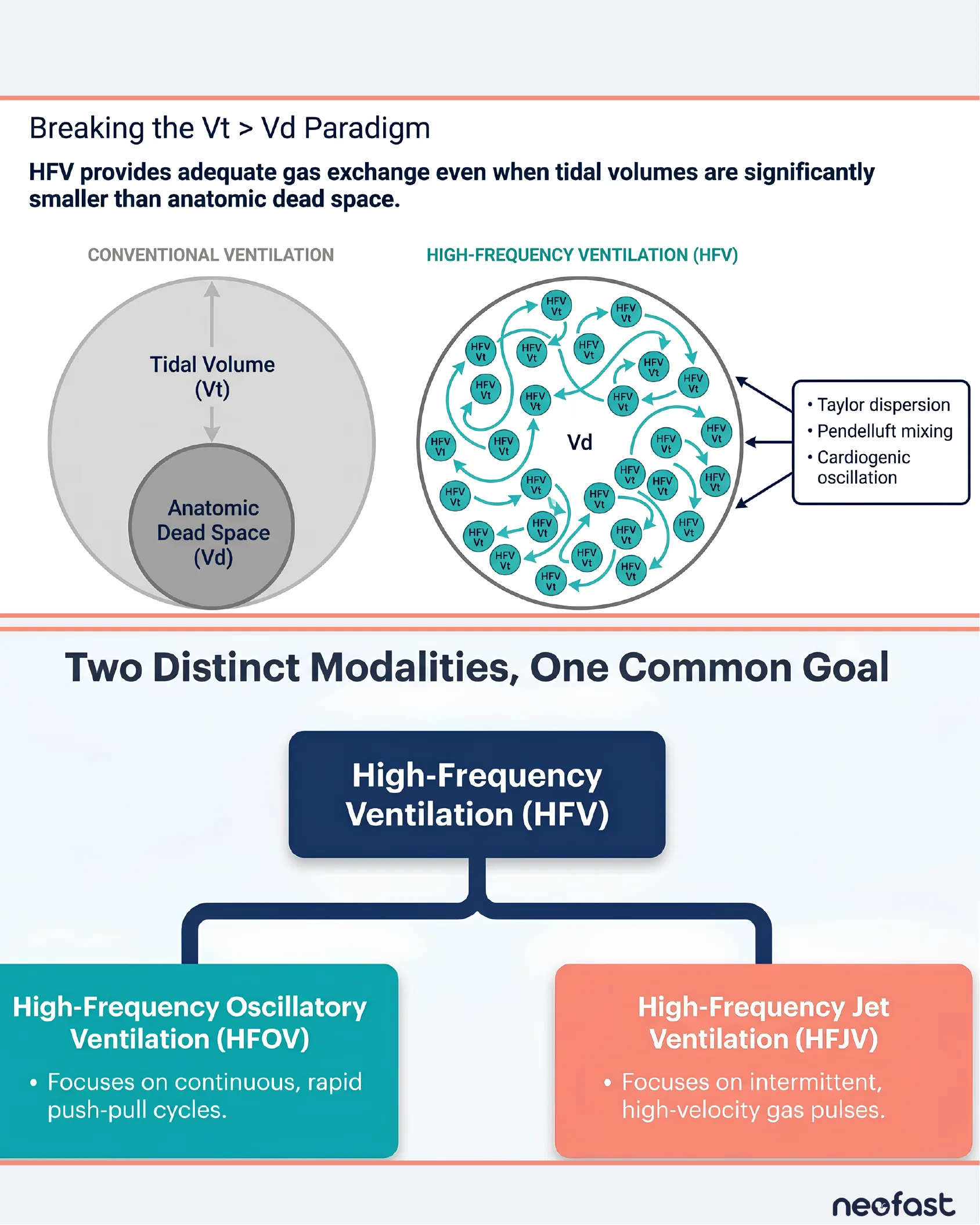
Breaking the Vt > Vd Paradigm
Breaking the Vt > Vd Paradigm
HFV provides adequate gas exchange even when tidal volumes are significantly smaller than anatomic dead space.
- Conventional Ventilation: Tidal Volume (Vt) is larger than Anatomic Dead Space (Vd).
- High-Frequency Ventilation (HFV): Multiple small HFV Vt packets circulate within and around the dead space (Vd) via specialized gas transport mechanisms:
- Taylor dispersion
- Pendelluft mixing
- Cardiogenic oscillation
Two Distinct Modalities, One Common Goal
- High-Frequency Ventilation (HFV) branches into two modalities:
- High-Frequency Oscillatory Ventilation (HFOV): Focuses on continuous, rapid push-pull cycles.
- High-Frequency Jet Ventilation (HFJV): Focuses on intermittent, high-velocity gas pulses.
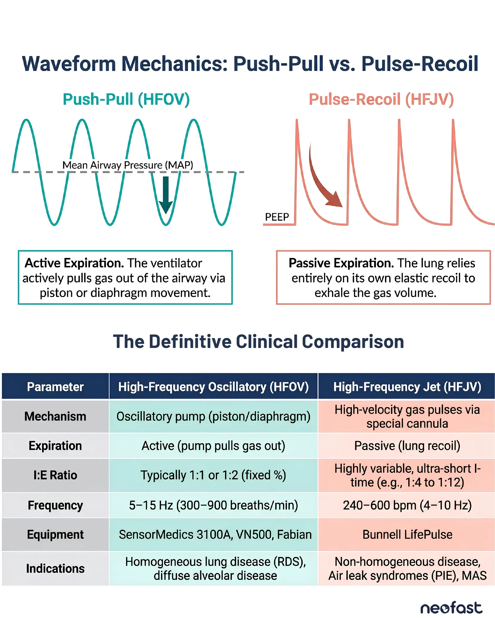
Waveform Mechanics: Push-Pull vs. Pulse-Recoil
Waveform Mechanics: Push-Pull vs. Pulse-Recoil
- Push-Pull (HFOV): Sinusoidal waveform oscillating around Mean Airway Pressure (MAP). Active Expiration – the ventilator actively pulls gas out of the airway via piston or diaphragm movement.
- Pulse-Recoil (HFJV): Sharp pulses rising from PEEP baseline. Passive Expiration – the lung relies entirely on its own elastic recoil to exhale the gas volume.
The Definitive Clinical Comparison
| Parameter | High-Frequency Oscillatory (HFOV) | High-Frequency Jet (HFJV) |
|---|---|---|
| Mechanism | Oscillatory pump (piston/diaphragm) | High-velocity gas pulses via special cannula |
| Expiration | Active (pump pulls gas out) | Passive (lung recoil) |
| I:E Ratio | Typically 1:1 or 1:2 (fixed %) | Highly variable, ultra-short I-time (e.g., 1:4 to 1:12) |
| Frequency | 5-15 Hz (300-900 breaths/min) | 240-600 bpm (4-10 Hz) |
| Equipment | SensorMedics 3100A, VN500, Fabian | Bunnell LifePulse |
| Indications | Homogeneous lung disease (RDS), diffuse alveolar disease | Non-homogeneous disease, Air leak syndromes (PIE), MAS |
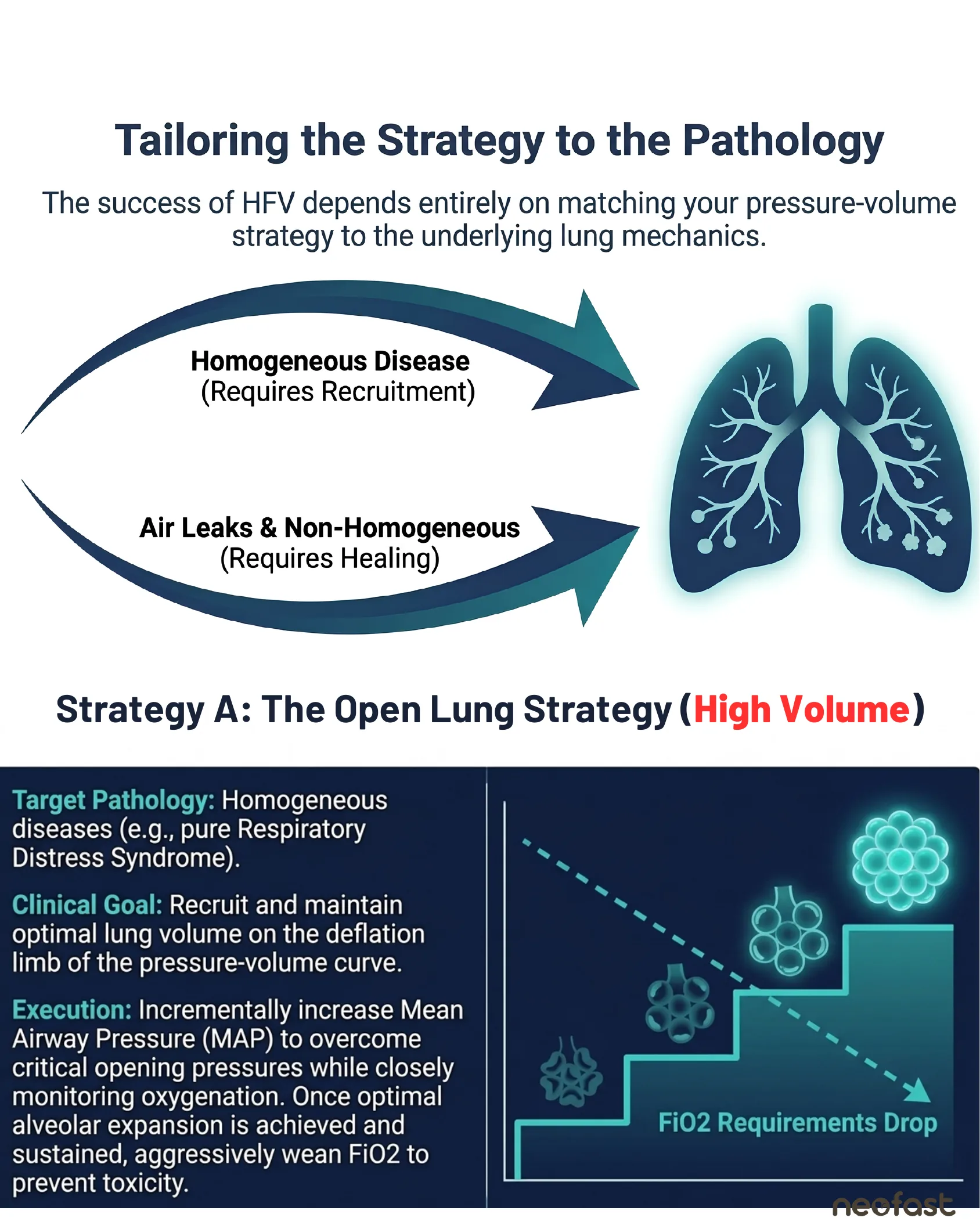
Tailoring the Strategy to the Pathology
Tailoring the Strategy to the Pathology
The success of HFV depends entirely on matching your pressure-volume strategy to the underlying lung mechanics.
- Homogeneous Disease (Requires Recruitment)
- Air Leaks & Non-Homogeneous (Requires Healing)
Strategy A: The Open Lung Strategy (High Volume)
Target Pathology: Homogeneous diseases (e.g., pure Respiratory Distress Syndrome).
Clinical Goal: Recruit and maintain optimal lung volume on the deflation limb of the pressure-volume curve.
Execution: Incrementally increase Mean Airway Pressure (MAP) to overcome critical opening pressures while closely monitoring oxygenation. Once optimal alveolar expansion is achieved and sustained, aggressively wean FiO2 to prevent toxicity.
FiO2 Requirements Drop (as alveolar recruitment improves in a stepwise fashion).
Strategy B: The Low PEEP Strategy
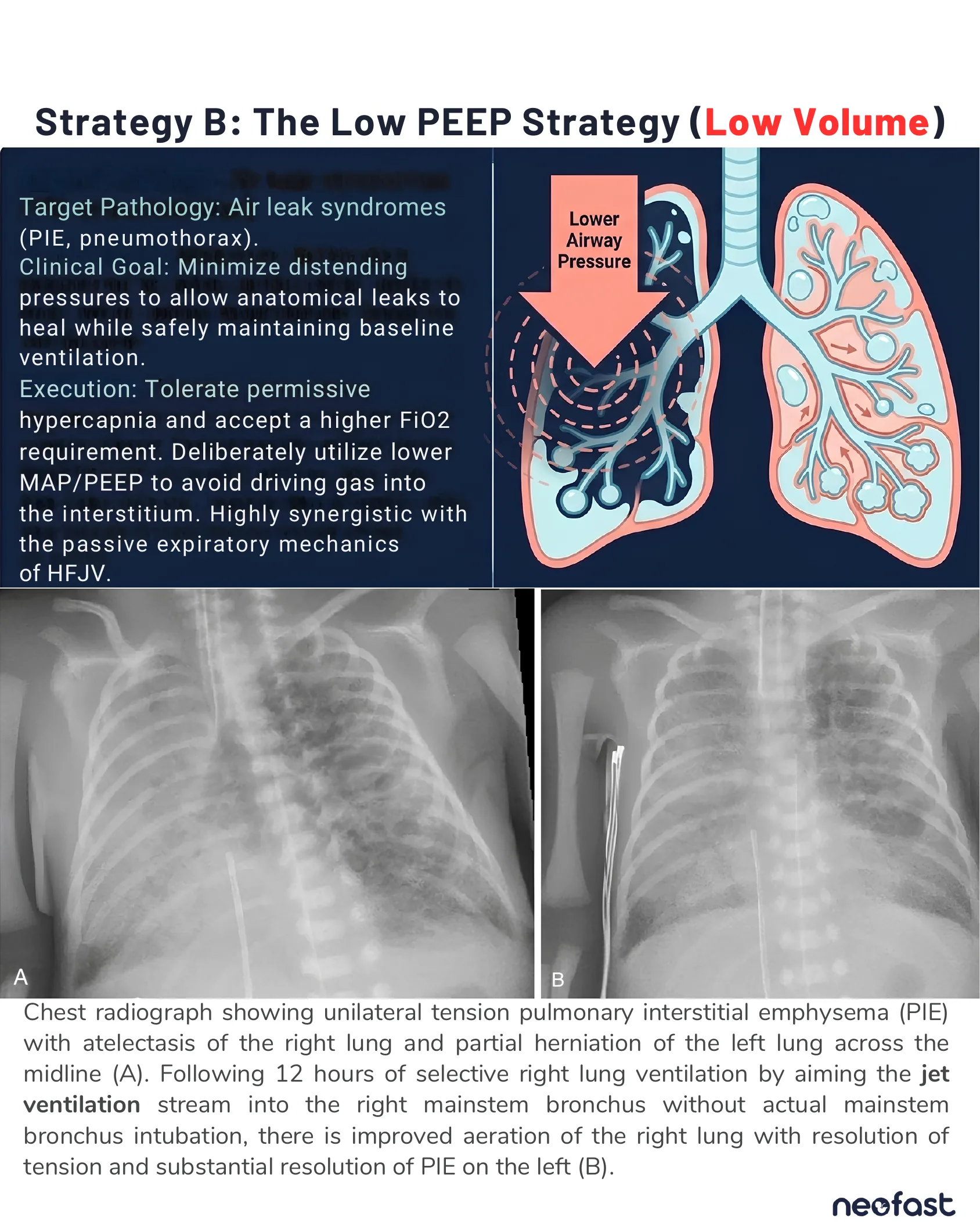
Strategy B: The Low PEEP Strategy (Low Volume)
Target Pathology: Air leak syndromes (PIE, pneumothorax).
Clinical Goal: Minimize distending pressures to allow anatomical leaks to heal while safely maintaining baseline ventilation.
Execution: Tolerate permissive hypercapnia and accept a higher FiO2 requirement. Deliberately utilize lower MAP/PEEP to avoid driving gas into the interstitium. Highly synergistic with the passive expiratory mechanics of HFJV.
Chest radiograph showing unilateral tension pulmonary interstitial emphysema (PIE) with atelectasis of the right lung and partial herniation of the left lung across the midline (A). Following 12 hours of selective right lung ventilation by aiming the jet ventilation stream into the right mainstem bronchus without actual mainstem bronchus intubation, there is improved aeration of the right lung with resolution of tension and substantial resolution of PIE on the left (B).
Radiographic Targets and Precision Monitoring
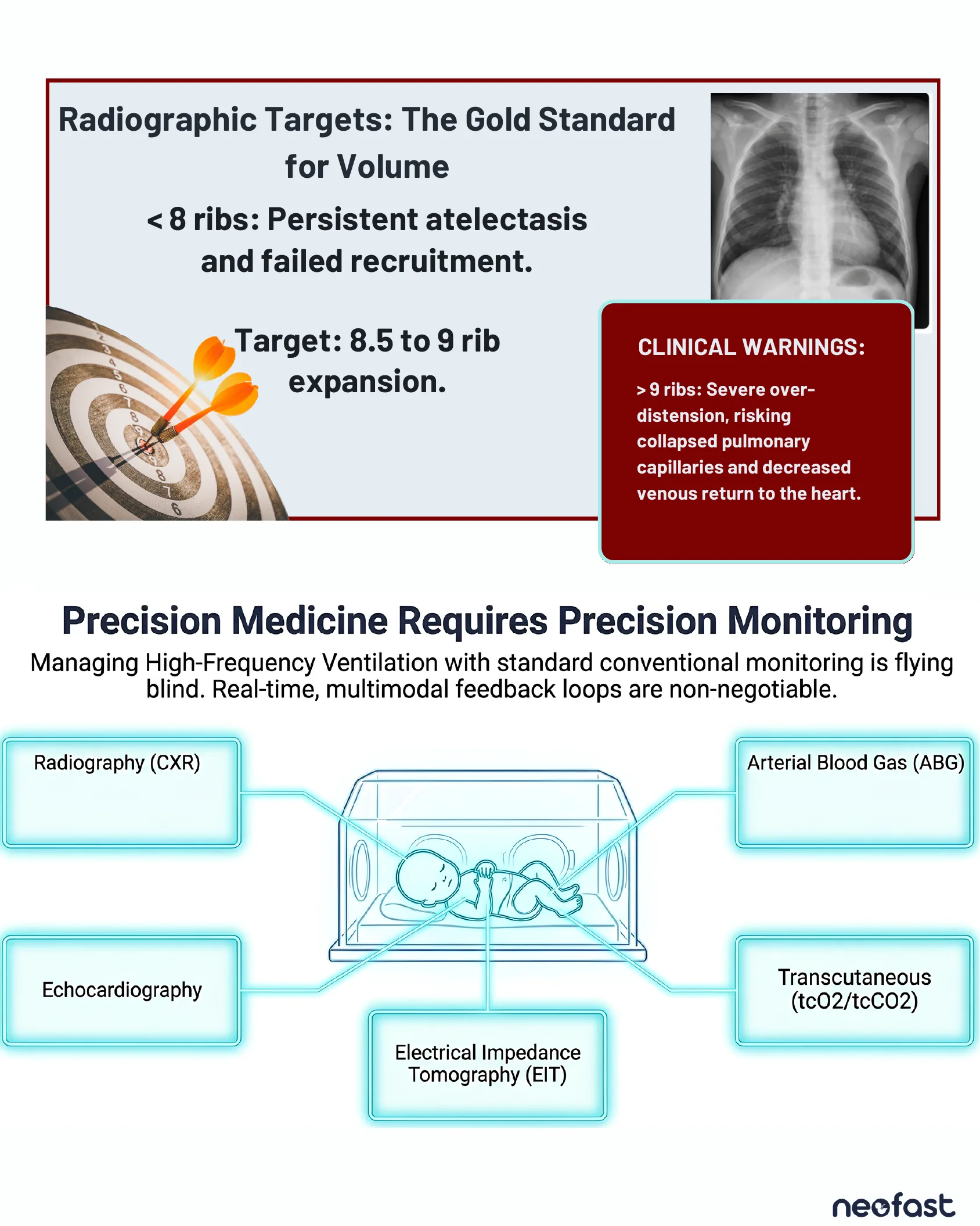
Radiographic Targets: The Gold Standard for Volume
- < 8 ribs: Persistent atelectasis and failed recruitment.
- Target: 8.5 to 9 rib expansion.
CLINICAL WARNINGS: > 9 ribs: Severe over-distension, risking collapsed pulmonary capillaries and decreased venous return to the heart.
Precision Medicine Requires Precision Monitoring
Managing High-Frequency Ventilation with standard conventional monitoring is flying blind. Real-time, multimodal feedback loops are non-negotiable.
- Radiography (CXR)
- Arterial Blood Gas (ABG)
- Echocardiography
- Electrical Impedance Tomography (EIT)
- Transcutaneous (tcO2/tcCO2)
Absolute Baseline vs. Dynamic Trend Monitoring
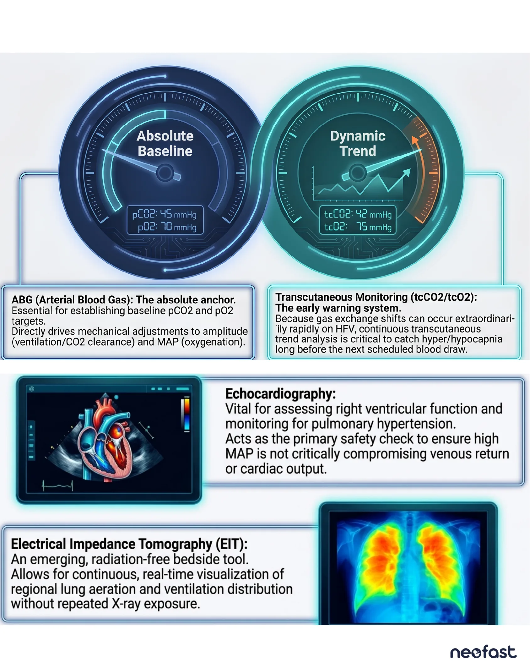
- Absolute Baseline (ABG – Arterial Blood Gas): pCO2: 45 mmHg, pO2: 70 mmHg. The absolute anchor. Essential for establishing baseline pCO2 and pO2 targets. Directly drives mechanical adjustments to amplitude (ventilation/CO2 clearance) and MAP (oxygenation).
- Dynamic Trend (Transcutaneous Monitoring – tcCO2/tcO2): tcCO2: 42 mmHg, tcO2: 75 mmHg. The early warning system. Because gas exchange shifts can occur extraordinarily rapidly on HFV, continuous transcutaneous trend analysis is critical to catch hyper/hypocapnia long before the next scheduled blood draw.
Echocardiography: Vital for assessing right ventricular function and monitoring for pulmonary hypertension. Acts as the primary safety check to ensure high MAP is not critically compromising venous return or cardiac output.
Electrical Impedance Tomography (EIT): An emerging, radiation-free bedside tool. Allows for continuous, real-time visualization of regional lung aeration and ventilation distribution without repeated X-ray exposure.
Basic Weaning Strategies and Clinical Synthesis
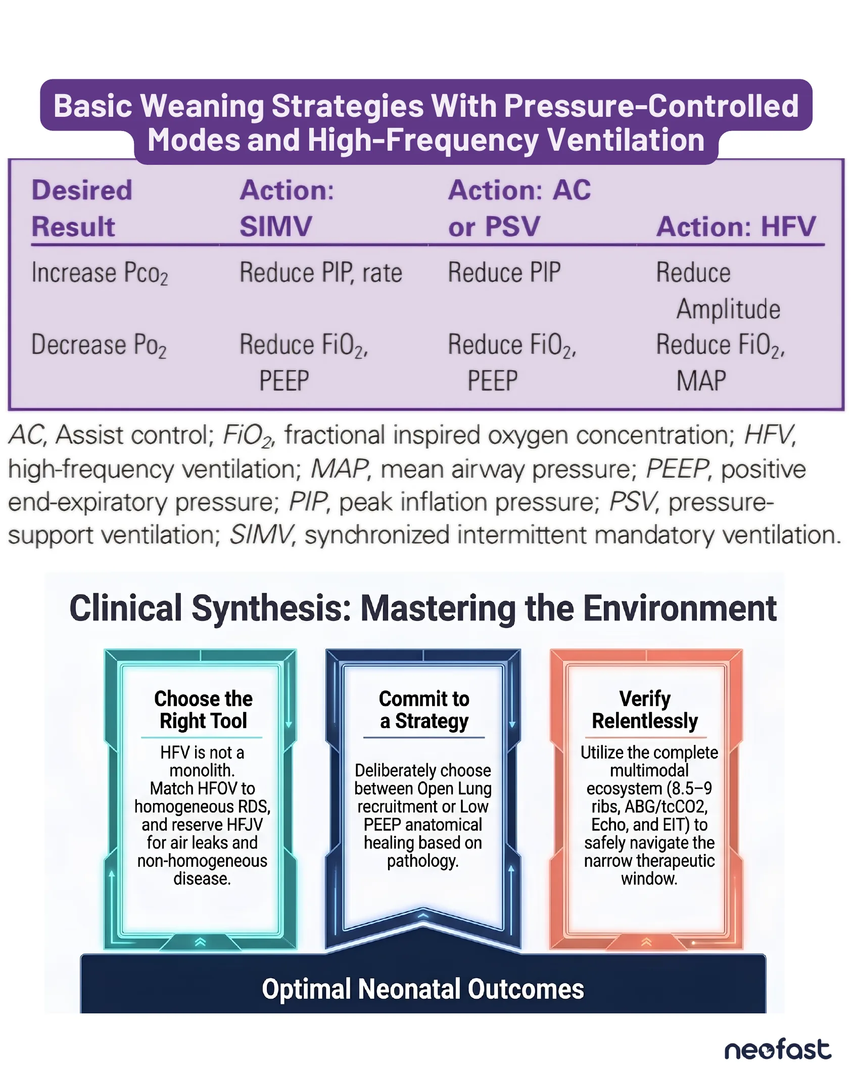
Basic Weaning Strategies With Pressure-Controlled Modes and High-Frequency Ventilation
| Desired Result | Action: SIMV | Action: AC or PSV | Action: HFV |
|---|---|---|---|
| Increase Pco2 | Reduce PIP, rate | Reduce PIP | Reduce Amplitude |
| Decrease Po2 | Reduce FiO2, PEEP | Reduce FiO2, PEEP | Reduce FiO2, MAP |
AC, Assist control; FiO2, fractional inspired oxygen concentration; HFV, high-frequency ventilation; MAP, mean airway pressure; PEEP, positive end-expiratory pressure; PIP, peak inflation pressure; PSV, pressure-support ventilation; SIMV, synchronized intermittent mandatory ventilation.
Clinical Synthesis: Mastering the Environment
- Choose the Right Tool: HFV is not a monolith. Match HFOV to homogeneous RDS, and reserve HFJV for air leaks and non-homogeneous disease.
- Commit to a Strategy: Deliberately choose between Open Lung recruitment or Low PEEP anatomical healing based on pathology.
- Verify Relentlessly: Utilize the complete multimodal ecosystem (8.5-9 ribs, ABG/tcCO2, Echo, and EIT) to safely navigate the narrow therapeutic window.
Optimal Neonatal Outcomes
Ventilatory Settings at Which Extubation Should Be Considered in Infants 2 Weeks of Age and Under
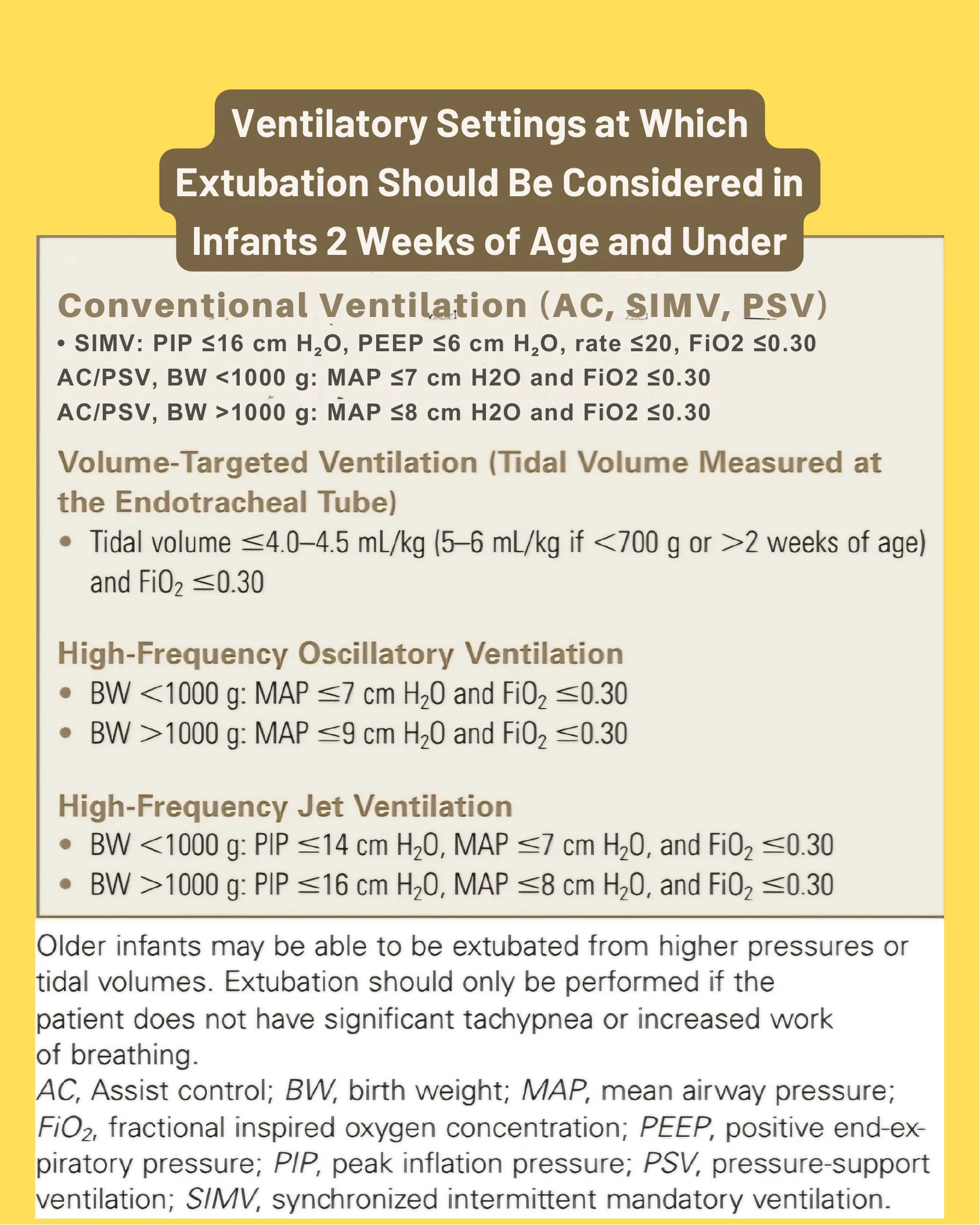
Conventional Ventilation (AC, SIMV, PSV)
- SIMV: PIP ≤16 cm H2O, PEEP ≤6 cm H2O, rate ≤20, FiO2 ≤0.30
- AC/PSV, BW <1000 g: MAP ≤7 cm H2O and FiO2 ≤0.30
- AC/PSV, BW >1000 g: MAP ≤8 cm H2O and FiO2 ≤0.30
Volume-Targeted Ventilation (Tidal Volume Measured at the Endotracheal Tube)
- Tidal volume ≤4.0–4.5 mL/kg (5–6 mL/kg if <700 g or >2 weeks of age) and FiO2 ≤0.30
High-Frequency Oscillatory Ventilation
- BW <1000 g: MAP ≤7 cm H2O and FiO2 ≤0.30
- BW >1000 g: MAP ≤9 cm H2O and FiO2 ≤0.30
High-Frequency Jet Ventilation
- BW <1000 g: PIP ≤14 cm H2O, MAP ≤7 cm H2O, and FiO2 ≤0.30
- BW >1000 g: PIP ≤16 cm H2O, MAP ≤8 cm H2O, and FiO2 ≤0.30
Older infants may be able to be extubated from higher pressures or tidal volumes. Extubation should only be performed if the patient does not have significant tachypnea or increased work of breathing.
AC, Assist control; BW, birth weight; MAP, mean airway pressure; FiO2, fractional inspired oxygen concentration; PEEP, positive end-expiratory pressure; PIP, peak inflation pressure; PSV, pressure-support ventilation; SIMV, synchronized intermittent mandatory ventilation.

neofast
- Venous hydration
- Continuous medication
- Medicines
- Intubation
- Other calculations and scores
- Procedures
- Respiratory and hemodynamic monitoring
- Growth and Development
Download on the App Store
Get it on Google Play
Frequently asked questions
What is the difference between HFOV and HFJV mechanics?
HFOV uses a sinusoidal push-pull waveform with active expiration (piston/diaphragm pulls gas out) at 5-15 Hz (300-900 breaths/min) and I:E typically 1:1 or 1:2; HFJV uses sharp pulse-recoil waveforms with passive expiration (lung recoil only) at 4-10 Hz (240-600 bpm) with ultra-short I-time (e.g., 1:4 to 1:12).
When should I choose HFOV versus HFJV?
HFOV is indicated for homogeneous lung disease such as pure RDS and diffuse alveolar disease; HFJV is reserved for non-homogeneous disease and air leak syndromes such as PIE and MAS.
What is the target rib expansion on chest X-ray during high-frequency ventilation?
The target is 8.5 to 9 rib expansion. Fewer than 8 ribs indicates persistent atelectasis and failed recruitment, while more than 9 ribs indicates severe over-distension risking collapsed pulmonary capillaries and decreased venous return to the heart.
What are the ABG and transcutaneous monitoring targets on HFV?
Baseline ABG targets are pCO2 45 mmHg and pO2 70 mmHg, driving amplitude and MAP adjustments. Transcutaneous trend targets are tcCO2 42 mmHg and tcO2 75 mmHg, serving as an early warning system for rapid gas exchange shifts between blood draws.
What are the extubation criteria from HFOV and HFJV in infants under 2 weeks old?
HFOV: BW <1000 g needs MAP ≤7 cm H2O and FiO2 ≤0.30; BW >1000 g needs MAP ≤9 cm H2O and FiO2 ≤0.30. HFJV: BW <1000 g needs PIP ≤14 cm H2O, MAP ≤7 cm H2O, FiO2 ≤0.30; BW >1000 g needs PIP ≤16 cm H2O, MAP ≤8 cm H2O, FiO2 ≤0.30.